The Striatum

The striatum is easily recognizable as striped. It is considered the input area of the basal ganglia, where the complex interconnection of a targeted movement begins. The front area also contains an important structure of the reward system.
Scientific support: Prof. Dr. Horst-Werner Korf
Published: 28.11.2025
Difficulty: intermediate
The striatum is easily recognizable as striped. It is considered the input area of the basal ganglia, where the complex interconnection of a targeted movement begins. The front area also contains an important structure of the reward system.
The corpus striatum – also known as the striatum and in German striped body – is the uppermost part of the basal ganglia and is one of the highly complex motor control circuits of the cerebrum. There, it primarily has an inhibitory effect, yet leads to excitation. And at least in its front, frontal part, it is indeed impressively striped.
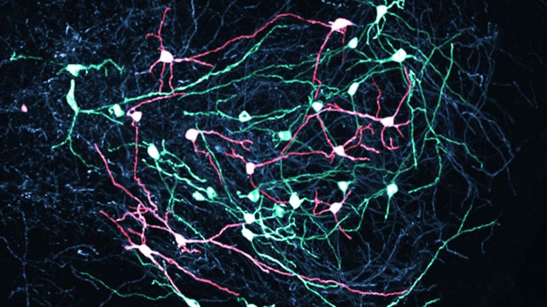
Related nuclei
This eponymous striping is caused by fiber tracts of the internal capsule, which partially traverse the striatum on their way between the thalamus and the cortex – and divide it into the caudate nucleus and the putamen. Originally, those two formed a single unit, both in embryonic development and in phylogenetic history. The internal capsule was added later, as the cortex became increasingly complex. And so, in animals that have poorly developed cortices and therefore no internal capsule, the two divisions of the striatum cannot be differentiated – or only with difficulty. In fact, even in humans, the putamen and caudate cannot be distinguished in terms of their cell types, function, and connections.
Circuits of motor regulation
The striatum regulates voluntary motor function; it is the “input element” in the complex system of the basal ganglia. What does this mean, for example, when reaching for a glass? Well, the striatum receives its inputs from neurons in the motor centers of the cortex, where an action plan is formed. The axons of these cortical neurons use glutamate as a neurotransmitter and thus exert an excitatory influence on the nerve cells of the striatum. To simplify, one could say that it integrates the motor “intention” of the cortex – the reaching movement – and “collects” it before it is actually executed.
The nerve cells of the striatum that are excited in this way are the medium spiny neurons (MSNs), named after the fine structures on their dendrites. And this is where the excitatory fibers from the cortex end. But the glass is not yet reached for. The MSNs themselves are inhibitory nerve cells. Their axons mainly extend to two other core areas: the substantia nigra, the black nucleus, and the globus pallidus, the pale nucleus, where they release GABA, an inhibitory neurotransmitter.
That sounds complicated. And it gets more complicated, because the substantia nigra sends its axons back into the striatum. These axons use dopamine as a neurotransmitter, which in turn has an inhibitory effect on the spiny cells. However, the inhibition of an inhibition is an excitation. In other words, the striatum–substantia nigra–striatum loop ultimately acts as a positive feedback loop and thus as an activator of the spiny cells.


When inhibition fails
Couldn't it be simpler? Apparently not, because we can see what happens without this positive feedback loop through the substantia nigra when its neurons die and no more dopamine reaches the striatum. The result is impaired motor function, or more precisely, “hypokinesia”: the body's movements become “small,” the steps “trippy,” the face “mask-like,” and, in general, there is a lack of movement. All these symptoms are seen in Parkinson's disease, which, among many other things, also destroys the dopaminergic cells of the substantia nigra. This affects the reaching for a glass in such a way that the movement towards it is slowed down and the hand trembles in a characteristic manner. Once the target is reached, drinking can proceed normally.
The signals “calculated” in the loop between the substantia nigra and the striatum are transmitted to the motor system via the globus pallidus, the other target nucleus of the striatum. Put simply, the globus pallidus is responsible for the “output” of the entire basal ganglia system.
Emotion and motor function
As mentioned above, the caudate nucleus and putamen appear to be separated. However, despite this separation, they meet at the front, lower part of the striatum and merge. This section is called the fundus striati (“bottom of the striatum”) or nucleus accumbens septi – septi because it is located near the septum.
The nucleus accumbens is characterized by intense inputs from the limbic system and thus represents a link between emotion and motor function. It is considered part of the reward system and has been referred to in the media as the “G-spot” of the brain because of its activation by food, some addictive drugs, and even money. That is a bit of an exaggeration. However, it is safe to say that the dopamine systems of motivation and motor function overlap in the nucleus accumbens – and that this intersection often contains things that make life worth living.
First published on August 23, 2011
Last updated on November 28, 2025