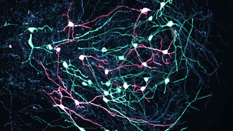
The Basal Ganglia

They are called caudate nucleus and putamen, are “torn” by fiber tracts, and lie deep within the brain. Every movement we perform deliberately is the result of their extensive networking, their mutual inhibition, and excitation.
Scientific support: Prof. Dr. Horst-Werner Korf
Published: 23.08.2011
Difficulty: intermediate
The Basal ganglia are non-cortical Gray matter of the Cerebrum whose main function is the global regulation of voluntary motor activity. As a student of anatomy, you should know: caudate + Putamen = striatum, putamen + Pallidum = lentiform nucleus.
Basal ganglia
Nuclei basales
The basal ganglia are a group of subcortical nuclei (located beneath the cerebral cortex) in the telencephalon. The basal ganglia include the globus pallidus and the striatum, and, depending on the author, other structures such as the substantia nigra and the subthalamic nucleus. The basal ganglia are primarily associated with voluntary motor function, but they also influence motivation, learning, and emotion.
Gray matter
Grey matter refers to a collection of nerve cell bodies, such as those found in nuclei or in the cortex.
Cerebrum
telencephalon
The cerebrum comprises the cerebral cortex (gray matter), the nerve fibers (white matter), and the basal ganglia. It is the largest part of the brain. The cortex can be divided into four cortical areas: the temporal lobe, frontal lobe, occipital lobe, and parietal lobe.
Its functions include the coordination of perception, motivation, learning, and thinking.
Putamen
A nucleus of the basal ganglia that, together with the caudate nucleus, forms the striatum. As part of the extrapyramidal motor system, it is involved in voluntary motor function (intentional movement).
Pallidum
globus pallidus
The globus pallidus, also known as the pallidum, is an important nucleus of the basal ganglia. It is a motor nucleus of the extrapyramidal system involved in the regulation of movement. The pallidum has an inhibitory and an excitatory part. The Latin name pallidus – pale – refers to the color of this nucleus.
Nucleus
In cell biology, the nucleus in a cell is the cell nucleus, which contains the chromosomes, among other things. In neuroanatomy, the nucleus in the nervous system refers to a collection of cell bodies – known as gray matter in the central nervous system and ganglia in the peripheral nervous system.
The brain has Gray matter and white matter The white matter consists mainly of nerve fibers. Such a nerve fiber typically consists of the Axon of the nerve cell, which is surrounded by special Glial cells called Oligodendrocytes. The largest fiber connection in the brain is the Corpus callosum between the two hemispheres. The gray matter consists of the cell bodies of the nerve cells, the well-known little gray cells – which, however, are by no means gray in the living brain, but rather pink. In addition, many special glial cells (mainly astrocytes) are also found in the gray matter. The gray matter occurs in the brain in layers, cortical, or in clusters as nuclei.
Gray matter
Grey matter refers to a collection of nerve cell bodies, such as those found in nuclei or in the cortex.
white matter
The white matter refers to the myelinated fibers of the nervous system that connect one neuron to another. The white color is caused by the myelin sheath surrounding the fibers.
Axon
axon
The axon is the extension of the nerve cell that is responsible for conducting nerve impulses to the next cell. An axon can branch out many times, reaching a large number of downstream nerve cells. It can be more than a meter long. The axon ends in one or more synapses.
Glial cells
Glia cells are the second largest group of cells in the brain after neurons. For a long time, they were considered inactive elements of the brain, referred to as "nerve cement." Today, we know that the different types of glia cells (astrocytes, oligodendrocytes, and microglia in the CNS; Schwann cells in the PNS) perform clearly defined tasks in the nervous system. For example, they respond to pathogens, play an important role in nourishing nerve cells, and insulate nerve fibers. They account for slightly more than 50 percent of the brain's cells, compared to neurons.
Oligodendrocytes
Cells of the central nervous system that form the myelin sheath around nerve cells, thereby increasing their conduction velocity. They belong to the glial cells.
Corpus callosum
As the largest commissure (connection in the brain), the corpus callosum connects the two cerebral hemispheres. It consists of 200-250 million nerve fibers and serves to exchange information.
The Gray matter of the telencephalon can be roughly divided as follows: On the one hand, there is the Cortex (literally: the “bark”), which lies just below the convoluted and folded surface of the telencephalon. And on the other hand, there are large, lumpy aggregates located in the center, at the base of the telencephalon, very close to the lateral ventricles. The Basal ganglia belong to these “non-cortical” gray masses.
Gray matter
Grey matter refers to a collection of nerve cell bodies, such as those found in nuclei or in the cortex.
Cortex
cortex cerebri
Cortex refers to a collection of neurons, typically in the form of a thin surface. However, it usually refers to the cerebral cortex, the outermost layer of the cerebrum. It is 2.5 mm to 5 mm thick and rich in nerve cells. The cerebral cortex is heavily folded, comparable to a handkerchief in a cup. This creates numerous convolutions (gyri), fissures (fissurae), and sulci. Unfolded, the surface area of the cortex is approximately 1,800cm².
lateral
A positional term – lateral means "towards the side." In relation to the nervous system, it refers to a direction at right angles to the neural axis, i.e., to the right or left.
Basal ganglia
Nuclei basales
The basal ganglia are a group of subcortical nuclei (located beneath the cerebral cortex) in the telencephalon. The basal ganglia include the globus pallidus and the striatum, and, depending on the author, other structures such as the substantia nigra and the subthalamic nucleus. The basal ganglia are primarily associated with voluntary motor function, but they also influence motivation, learning, and emotion.
What are they – and what are they not?
The collective term “basal ganglia” is not clearly defined and has changed over time. Some regions that were previously included under this term have now been excluded for functional reasons, as they are not directly part of the motor system. This is because the regulation of voluntary motor function and motor Memory are the core tasks of the basal ganglia.
It has proven useful to approach the Basal ganglia using a kind of “historical exclusion process.” For a long time, the gray masses flanking the third ventricle of the Diencephalon on the right and left sides are not considered being part of the basal ganglia: These are the two thalami. The amygdalae, the almond-shaped nuclei located on both sides inside and above the Temporal lobe and in front of the pes hippocampi (like a soccer ball in front of a soccer shoe, i.e., the pes hippocampi), are also no longer considered part of the basal ganglia. Most experts now assign them to the Limbic system because they serve the affective rather than the motor system.
In addition, the claustrum is no longer considered part of the basal ganglia. This is the layer of Gray matter located just below the Cortex of the Insula (where Nobel Prize winner Francis Crick believed consciousness to be located). Current research suggests that the claustrum is a piece of “scattered cortex” that may be involved in cognitive functions, desires, and addictions.
This leaves the motor basal ganglia in the narrower, modern sense, namely the Caudate nucleus (the “tail core”) and the Putamen (the “shell core”). The caudate nucleus, the “tailed nucleus,” has a very peculiar, curved shape. It follows the curved contour of the lateral ventricle, to which the caudate Nucleus is directly adjacent. Particularly in its thick front section, known as the head or caput nuclei caudati, the caudate nucleus is connected to the putamen by many bridges of gray matter.
In fact, both were originally – both in terms of phylogenetic development and embryonic development – a single core area. It was only “torn apart” by a massive bundle of white matter that pushed between them. This bundle of fibers is the internal capsule, in which the cortical efferents descending to other regions, but also the reciprocal thalamocortical fiber connections, are bundled. When viewed together, the gray bridges mentioned above and the white fiber bundles of the internal capsule that squeeze in between them actually form a gray-white striped pattern in the sectional view – especially in the anterior part of the basal ganglia, which lies between the putamen and the head of the caudate nucleus. This has earned them the name “striatum.”
At the very front and very bottom, the head of the caudate nucleus and the base of the putamen are actually still completely undivided and connected by a broad bridge of gray substance. This is the Nucleus accumbens septi, also known as the ventral Striatum or fundus striati.
Finally, there is the globus pallidus, which literally means “pale sphere.” It is often simply referred to as the Pallidum. The pallidum is pale because its nerve cells – unlike those of the other basal ganglia – contain very little pigment (neuromelanin and lipofuscin). The pallidum is located close to the putamen, toward the center, toward the diencephalon. Together, the putamen and pallidum are also referred to as the “lentiform nucleus.” This term, which is based on their purely macroscopic appearance, should no longer be used today, as it combines sections that are completely different in terms of their developmental history and function. Here, too, the internal capsule has done its disruptive work: the pallidum originally belonged to the diencephalon, in the ventral thalamus, through which the internal capsule also grew. It thus pushed the pallidum to the side, toward the putamen. What remained of the ventral thalamus in its original location is now called the subthalamic nucleus.
Memory
Memory is a generic term for all types of information storage in the organism. In addition to pure retention, this also includes the absorption of information, its organization, and retrieval.
Basal ganglia
Nuclei basales
The basal ganglia are a group of subcortical nuclei (located beneath the cerebral cortex) in the telencephalon. The basal ganglia include the globus pallidus and the striatum, and, depending on the author, other structures such as the substantia nigra and the subthalamic nucleus. The basal ganglia are primarily associated with voluntary motor function, but they also influence motivation, learning, and emotion.
Diencephalon
The diencephalon (midbrain) includes the thalamus and hypothalamus, among other structures. Together with the cerebrum, it forms the forebrain. The diencephalon contains centers for sensory perception, emotion, and the control of vital functions such as hunger and thirst.
Temporal lobe
Lobus temporalis
The temporal lobe is one of the four lobes of the cerebrum and is located laterally (on the side) at the bottom. It contains important areas such as the auditory cortex and parts of Wernicke's area, as well as areas for higher visual processing; deep within it lies the medial temporal lobe with structures such as the hippocampus.
Limbic system
The limbic system is a functional unit in the brain. It consists of interconnected structures, primarily in the cerebrum and diencephalon. The structures assigned to the system vary depending on the source, but the most important components are the hippocampus, amygdala, cingulate gyrus, septum, and mammillary bodies. The limbic system is involved in autonomic and visceral processes as well as in mechanisms of emotion, memory, and learning. Some authors mistakenly reduce the limbic system to the emotional world by referring to it as the "emotional brain."
Gray matter
Grey matter refers to a collection of nerve cell bodies, such as those found in nuclei or in the cortex.
Cortex
cortex cerebri
Cortex refers to a collection of neurons, typically in the form of a thin surface. However, it usually refers to the cerebral cortex, the outermost layer of the cerebrum. It is 2.5 mm to 5 mm thick and rich in nerve cells. The cerebral cortex is heavily folded, comparable to a handkerchief in a cup. This creates numerous convolutions (gyri), fissures (fissurae), and sulci. Unfolded, the surface area of the cortex is approximately 1,800cm².
Insula
lobus insularis
The insula is a recessed part of the cortex (cerebral cortex) that is covered by the frontal, temporal, and parietal lobes. This overlay is called the opercula (lid). The insula influences the motor and sensory functions of the intestines and is considered to be the link between cognitive and emotional elements in pain processing. It is also involved in processes such as taste and physical self-awareness.
Caudate nucleus
nucleus caudatus
Part of the basal ganglia, it forms the striatum together with the putamen. Anatomically, the caudate nucleus is located frontally in the center of the brain and extends backward, forming a C shape. It consists of a head (caput nuclei caudati), a body (corpus nuclei caudati), and a tail (cauda nuclei caudati). In contrast to the more motor-related parts of the basal ganglia, this area is strongly connected to the prefrontal cortex in addition to its motor functions. As a result, this part of the striatum is also heavily involved in cognition, motivation, and emotion.
Putamen
A nucleus of the basal ganglia that, together with the caudate nucleus, forms the striatum. As part of the extrapyramidal motor system, it is involved in voluntary motor function (intentional movement).
lateral
A positional term – lateral means "towards the side." In relation to the nervous system, it refers to a direction at right angles to the neural axis, i.e., to the right or left.
Nucleus
In cell biology, the nucleus in a cell is the cell nucleus, which contains the chromosomes, among other things. In neuroanatomy, the nucleus in the nervous system refers to a collection of cell bodies – known as gray matter in the central nervous system and ganglia in the peripheral nervous system.
white matter
The white matter refers to the myelinated fibers of the nervous system that connect one neuron to another. The white color is caused by the myelin sheath surrounding the fibers.
Nucleus accumbens
The nucleus accumbens is a nucleus in the basal ganglia that receives dopaminergic (dopamine-responsive) inputs from the ventral tegmental area. It is associated with reward and attention, but also with addiction. In pain processing, it is involved in motivational aspects of pain (reward, pain reduction) and in the effect of placebos.
ventral
A positional term – ventral means "towards the abdomen." In relation to the nervous system, it refers to a direction perpendicular to the neural axis, i.e., downwards or forwards.
In animals (that do not walk upright), the term is simpler, as it always means toward the abdomen. Due to the upright posture of humans, the brain bends in relation to the spinal cord, making ventral mean "forward."
Striatum
Corpus striatum
The striatum is a central structure of the basal ganglia. It consists of the caudate nucleus and putamen; the nucleus accumbens is also functionally part of it as its ventral portion. As the most important input structure of the basal ganglia, the striatum plays an essential role in controlling movement sequences as well as in cognition, motivational processes, and the reward system.
Pallidum
globus pallidus
The globus pallidus, also known as the pallidum, is an important nucleus of the basal ganglia. It is a motor nucleus of the extrapyramidal system involved in the regulation of movement. The pallidum has an inhibitory and an excitatory part. The Latin name pallidus – pale – refers to the color of this nucleus.


Function
The ventral Striatum is part of our “reward system.” The nerve cells there are primarily activated when we expect or receive a reward or satisfaction. The ventral striatum therefore also plays an important role in all kinds of addictions and in both good and bad habits.
The remaining, much larger areas of the basal ganglia, i.e., the caudate, putamen, and pallidum, are responsible for motor function, or more precisely, its regulation. In particular, they serve to initiate, i.e., actually start, and terminate voluntary motor acts. They also contain part of our motor Memory. These are motor skills such as running, riding a bike, or playing the piano, which we acquire over the course of our lives.
In simple terms, it can be said that disturbances in the function of the Basal ganglia are associated with global motor deficits. It is not individual muscle groups or extremities that are affected. Rather, all muscles suffer from similar coordination problems. The “paradigmatic” basal ganglia disorder is Parkinson's disease, which is characterized by increased muscle tone, i.e., rigidity, lack of movement, slow and limited movements, and the well-known tremor (shaking).
It should be noted that the basal ganglia do not exert their regulatory influence on the motor system directly, but via a neural loop that connects them to the thalamus and the (motor) Cortex. When a movement is to be initiated, the cortex sends excitatory impulses via glutamatergic fibers down to the basal ganglia and into the Putamen and the caudate nucleus, which represent, so to speak, the “input” side of the basal ganglia. The shell and caudate nuclei in turn send inhibitory impulses to the Pallidum. Its fibers, which also use the inhibitory Neurotransmitter GABA, act as the “output” of the basal ganglia to a Nucleus in the Dorsal thalamus However, the Inhibition of an inhibition (disinhibition) is equivalent to excitation. Therefore, this thalamic nucleus becomes active when the cortex is active, as well. The thalamic nucleus (called the ventrolateral anterior nucleus) sends its fibers to those areas of the cortex whose nerve cells then send the actual motor pathways that descend to the motor neurons in the brain and spinal cord.
The well-known substantia nigra, the “black nucleus” of the midbrain, is also involved in the regulation of this “cortico-basal ganglionic-thalamo-cortical” loop. The basal ganglia send efferents there, partly directly and partly via the Subthalamic nucleus of the Diencephalon. The substantia nigra, in turn, sends dopaminergic axons, the nigrostriatal tract, to the striatum, whose neurons receive the Dopamine signals via presynaptic dopamine transporters (DaT) and postsynaptically located D2 receptors. When there is a dopamine deficiency, the expression of DaT is downregulated. If there is a lack of dopamine in the striatum – for example, in Parkinson's disease, because the nerve cells in the Substantia nigra have died – this results in the notorious rigidity and lack of movement. Conversely, for example in the case of damage to the subthalamic nucleus or the pallidum, uncontrolled and completely involuntary “movement outbreaks” of individual extremities or even the entire body can occur, which cannot be stopped at will.
Then there is Huntington's disease, a hereditary disorder of the central nervous system which, like Parkinson's disease, is a multisystem disorder of the central nervous system with “movement outbreaks.” It leads to the loss of neurons in the striatum and manifests macroscopically as a flattening of the Caudate nucleus with corresponding enlargement of the lateral ventricles. The cause is a mutation in the huntingtin gene, which leads to an increase in the base triplets CAG (cytosine, adenine, guanine) and is therefore referred to as a trinucleotide disorder.
ventral
A positional term – ventral means "towards the abdomen." In relation to the nervous system, it refers to a direction perpendicular to the neural axis, i.e., downwards or forwards.
In animals (that do not walk upright), the term is simpler, as it always means toward the abdomen. Due to the upright posture of humans, the brain bends in relation to the spinal cord, making ventral mean "forward."
Striatum
Corpus striatum
The striatum is a central structure of the basal ganglia. It consists of the caudate nucleus and putamen; the nucleus accumbens is also functionally part of it as its ventral portion. As the most important input structure of the basal ganglia, the striatum plays an essential role in controlling movement sequences as well as in cognition, motivational processes, and the reward system.
Memory
Memory is a generic term for all types of information storage in the organism. In addition to pure retention, this also includes the absorption of information, its organization, and retrieval.
Basal ganglia
Nuclei basales
The basal ganglia are a group of subcortical nuclei (located beneath the cerebral cortex) in the telencephalon. The basal ganglia include the globus pallidus and the striatum, and, depending on the author, other structures such as the substantia nigra and the subthalamic nucleus. The basal ganglia are primarily associated with voluntary motor function, but they also influence motivation, learning, and emotion.
Cortex
cortex cerebri
Cortex refers to a collection of neurons, typically in the form of a thin surface. However, it usually refers to the cerebral cortex, the outermost layer of the cerebrum. It is 2.5 mm to 5 mm thick and rich in nerve cells. The cerebral cortex is heavily folded, comparable to a handkerchief in a cup. This creates numerous convolutions (gyri), fissures (fissurae), and sulci. Unfolded, the surface area of the cortex is approximately 1,800cm².
excitatory
Exciting synapses are described as excitatory when they depolarize the subsequent cell membrane and can thus lead to the formation of an action potential. An excitatory effect is usually produced by an exciting transmitter (messenger substance), such as glutamate. The opposite is an inhibitory synapse.
Putamen
A nucleus of the basal ganglia that, together with the caudate nucleus, forms the striatum. As part of the extrapyramidal motor system, it is involved in voluntary motor function (intentional movement).
Pallidum
globus pallidus
The globus pallidus, also known as the pallidum, is an important nucleus of the basal ganglia. It is a motor nucleus of the extrapyramidal system involved in the regulation of movement. The pallidum has an inhibitory and an excitatory part. The Latin name pallidus – pale – refers to the color of this nucleus.
Neurotransmitter
A neurotransmitter is a chemical messenger, an intermediary substance. It is released by the sender neuron at the sites of cell-cell communication and has an excitatory or inhibitory effect on the receiver neuron.
Nucleus
In cell biology, the nucleus in a cell is the cell nucleus, which contains the chromosomes, among other things. In neuroanatomy, the nucleus in the nervous system refers to a collection of cell bodies – known as gray matter in the central nervous system and ganglia in the peripheral nervous system.
dorsal
The positional term dorsal means "towards the back." In relation to the nervous system, it refers to a direction perpendicular to the neural axis, i.e., upwards towards the head or backwards.
In animals that do not walk upright, the term is simpler, as it always means toward the back. Due to the upright posture of humans, the brain bends in relation to the spinal cord, making dorsal mean "upward."
Dorsal thalamus
Thalamus dorsals
The thalamus is the largest structure in the diencephalon and is located above the hypothalamus. The thalamus is considered the "gateway to consciousness" because its nuclei are the transit station for all information to the cortex (cerebral cortex) – except for olfactory information, which first reaches the olfactory areas of the brain directly. At the same time, they also receive massive cortical inputs so it might be better to regard this a thalami-cortical system. The nuclei of the thalamus are grouped together. The term "gateway to consciousness" also refers to attention control, sleep-wake regulation, and consciousness modulation by the intralaminar nuclei.
Inhibition
Neuronal inhibition describes the phenomenon whereby a sender neuron sends an impulse to a receiver neuron, causing the latter's activity to decrease. The most important inhibitory neurotransmitter is GABA.
Subthalamic nucleus
Nucleus subthalamicus
Although the subthalamic nucleus is a nucleus of the subthalamus in the diencephalon, it is functionally closely integrated into the motor control of the basal ganglia. It plays a role in impulse control, movement control, and inhibition of unwanted movements. Damage to this nucleus can lead to temporary, uncontrolled, jerky movements of the extremities – known as ballism. Doctors have already achieved successful treatment outcomes in both obsessive-compulsive disorder and Parkinson's disease by artificially stimulating this region with a neuroimplant.
Diencephalon
The diencephalon (midbrain) includes the thalamus and hypothalamus, among other structures. Together with the cerebrum, it forms the forebrain. The diencephalon contains centers for sensory perception, emotion, and the control of vital functions such as hunger and thirst.
Dopamine
Dopamine is an important neurotransmitter in the central nervous system that belongs to the catecholamine group. It plays a role in motor function, motivation, emotion, and cognitive processes. Disruptions in the function of this transmitter play a role in many brain disorders, such as schizophrenia, depression, Parkinson's disease, and substance dependence.
Substantia nigra
A nucleus complex in the ventral mesencephalon that plays a central role in initiating and modulating movement. It appears dark due to neuromelanin. Its dopaminergic neurons project via the nigrostriatal pathways to the putamen and caudate nucleus. Failure of these neurons leads to the typical symptoms of Parkinson's disease.
Caudate nucleus
nucleus caudatus
Part of the basal ganglia, it forms the striatum together with the putamen. Anatomically, the caudate nucleus is located frontally in the center of the brain and extends backward, forming a C shape. It consists of a head (caput nuclei caudati), a body (corpus nuclei caudati), and a tail (cauda nuclei caudati). In contrast to the more motor-related parts of the basal ganglia, this area is strongly connected to the prefrontal cortex in addition to its motor functions. As a result, this part of the striatum is also heavily involved in cognition, motivation, and emotion.
lateral
A positional term – lateral means "towards the side." In relation to the nervous system, it refers to a direction at right angles to the neural axis, i.e., to the right or left.