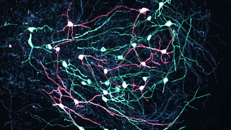
Senses in Motion
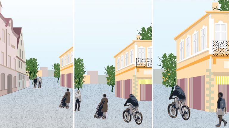
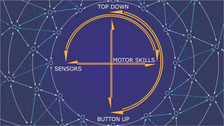
Stimulus – Processing – Response: We’re familiar with this chain from school. But in reality, sensory and motor systems work together very early on in the processing of sensory stimuli.
Scientific support: Prof. Dr. Julia Schiemann
Published: 21.12.2021
Difficulty: intermediate
- Stimuli from the environment activate the sensory organs; nerve cells send the information to the brain, which evaluates it and sends commands to the muscles, thereby triggering a movement.
- However, sensory and motor systems interact early on in the processing of stimuli.
- The brain constantly makes predictions about what input from the sensory organs – such as the eyes – will be like and compares this with the actual input. In this way, it calculates our own movements; our Perception of the world remains stable.
Perception
The term describes the complex process of gathering and processing information from stimuli in the environment and from the internal states of a living being. The brain combines the information, which is perceived partly consciously and partly unconsciously, into a subjectively meaningful overall impression. If the data it receives from the sensory organs is insufficient for this, it supplements it with empirical values. This can lead to misinterpretations and explains why we succumb to optical illusions or fall for magic tricks.
The Lateral geniculate nucleus (LGN) is located in the thalamus and is part of the Visual pathway Its neurons send visual information to the Visual cortex What is less well known is that there is also a feedback loop in the opposite direction. The Cortex thus appears to influence its own input and, for example, flexibly increase the response strength of the LGN. The fact is: movement can upregulate the thalamus. For neuroscientist Laura Busse of LMU Munich, this raises the question: Is the thalamus simply modulated by the fact that body movement upregulates the visual cortex, and is this modulation relayed to the thalamus via a feedback loop? To investigate this, she conducted experiments together with colleagues: Using optogenetic techniques, the team temporarily shut down the visual cortex in mice. Yet movement still upregulated the thalamus. The effect, therefore, cannot be due to the feedback loop alone. One possible explanation: Neurotransmitters that reach the thalamus directly could play a crucial role in upregulating the thalamus during movement.
lateral
A positional term – lateral means "towards the side." In relation to the nervous system, it refers to a direction at right angles to the neural axis, i.e., to the right or left.
Lateral geniculate nucleus
corpus geniculatum laterale
The lateral geniculate nucleus is the section of the thalamus (the largest part of the diencephalon) where around 90% of the optic nerve axons terminate. It has a characteristic stratification into six cell layers. The nerve cells of the lateral geniculate nucleus send their projections to the visual cortex. Together with the medial geniculate nucleus, it forms the metathalamus.
Visual pathway
The visual pathway refers to the network of nerve cells involved in visual perception. In mammals, it runs from the retinal ganglion cells in the eye – as the optic nerve to the optic chiasm, then as the visual tract – via the only switching point in the lateral geniculate nucleus to the primary visual cortex.
Visual cortex
The visual cortex refers to the areas of the occipital lobe that are involved in processing visual information. These include the primary visual cortex and the associative visual cortices V1 to V5. According to Brodmann, the visual cortex comprises areas 17, 18, and 19.
Cortex
cortex cerebri
Cortex refers to a collection of neurons, typically in the form of a thin surface. However, it usually refers to the cerebral cortex, the outermost layer of the cerebrum. It is 2.5 mm to 5 mm thick and rich in nerve cells. The cerebral cortex is heavily folded, comparable to a handkerchief in a cup. This creates numerous convolutions (gyri), fissures (fissurae), and sulci. Unfolded, the surface area of the cortex is approximately 1,800cm².
It seems so simple. In, out. A stimulus comes in, a response goes out. For example, if a ball flies straight toward us, nerve cells in the Retina of the Eye are activated. These cells transmit the information in the form of electrical signals to the brain, which then processes the information: the ball, its direction of flight, and its speed. It then sends commands to the muscles. In response, our arms and hands move, and we catch the ball.
This chain of processing separates the sensory system from the motor system and seems straightforward at first glance. But as is so often the case, the reality is more complicated. This is because both systems interact early on in neural processing. It’s not just the sensory system that influences the motor system; the motor system also influences the sensory system.
One thing becomes clear here: The brain doesn’t just react – it acts. It tries to look into the future. “Our brain is constantly making predictions about what input from the sensory organs – such as the eyes – will be like,” says neurobiologist Mark Hübener of the Max Planck Institute for Biological Intelligence in Martinsried. “Many neurons are busy comparing the expected sensory input with the actual input.” Essentially, this touches on a very old question that the eminent German physiologist and physicist Hermann von Helmholtz already posed in the 19th century: How is it that our image of the world remains stable and does not wobble, even though we are constantly moving our eyes and head, causing the image on the retina to shift constantly?
If you pan a camera back and forth, it results in a blurry image. And in principle, it should be the same with vision. But it isn’t. “The idea is this: The Visual system receives a copy of the movement commands that control the movement of the eyes and head,” says Hübener. Researchers refer to this as an efferent copy. “In this process, the sensory input expected based on the strength and direction of our eye and head movements is compared with the actual input,” explains the neurobiologist. If there is no difference, our image of the world remains stable. This effectively filters out our own movements from the visual input. Incidentally, it is possible to trick this system. If you gently press your eyelid with a finger to carefully move the eyeball back and forth, the world – which is actually static – suddenly appears to move.
To counteract eye or head movements, the brain also relies on compensatory movements. This is being researched by biologist Boris Chagnaud from the University of Graz. “The efferent copies inform the visual system when and how our head is moving, and the oculomotor system then compensates for these movements,” says Chagnaud. The oculomotor system specifically controls the movements of the eyes. “It moves the eyes in the opposite direction of the head, thereby keeping the gaze stable,” says the biologist. And the Spinal cord also plays a role. When walking, we always move slightly up and down, while simultaneously swaying a little from side to side. As a result, the position of the eyes also changes. “The spinal cord helps to compensate for these passive eye movements,” says Boris Chagnaud. It sends copies of the motor signals to the oculomotor system, among other systems. The idea here is always to keep the impact of movements on the sensory systems as low as possible.
Retina
The retina is the inner layer of the eye covered with pigment epithelium. The retina is characterized by an inverse (reversed) arrangement: light must first pass through several layers before it hits the photoreceptors (cones and rods). The signals from the photoreceptors are transmitted via the optic nerve to the processing areas of the brain. The reason for the inverse arrangement is the evolutionary development of the retina, which is a protrusion of the brain.
The retina is approximately 0.2 to 0.5 mm thick.
Eye
bulbus oculi
The eye is the sensory organ responsible for perceiving light stimuli – electromagnetic radiation within a specific frequency range. The light visible to humans lies in the range between 380 and 780 nanometers.
Intelligence
Intelligence
Collective term for human cognitive performance. According to British psychologist Charles Spearman, cognitive performance in different areas correlates with a general factor (g factor) of intelligence. This means that intelligence can be expressed as a single value. American psychologist Howard Gardner, among others, has developed a counter-concept to this, known as the "theory of multiple intelligences." According to this theory, intelligence develops independently in the following eight areas: linguistic, logical-mathematical, musical-rhythmic, visual-spatial, bodily-kinesthetic, naturalistic, intrapersonal, and interpersonal.
Visual system
The visual system is the part of the nervous system that processes visual information. It primarily comprises the eye, the optic nerve, the optic chiasm, the optic tract, the lateral geniculate nucleus, the optic radiation, the primary visual cortex, and the visual association cortices.
Spinal cord
medulla spinalis
The spinal cord is the part of the central nervous system located in the spine. It contains both the white matter of the nerve fibers and the gray matter of the cell nuclei. Simple reflexes such as the knee-jerk reflex are already processed here, as sensory and motor neurons are directly connected. The spinal cord is divided into the cervical, thoracic, lumbar, and sacral spinal cord.
Video instead of individual frames
In the process, our own nerve cells sometimes really trick us. This becomes evident when reading. We make purposeful Eye movements to focus on sections of a line. However, rapid eye movements – called saccades – don’t align well with sharp vision. Therefore, efferent copies inform the Visual system about the upcoming eye movements. “Even though we don’t notice it: During saccades, a part of the brain – the so-called frontal eye field – blocks out the moving visual information,” says Boris Chagnaud. We don’t notice this, so we don’t see individual frames, but rather, in a sense, a continuous film. “In this way, the brain constructs an impression of the environment that is not perceived by our eyes in this manner,” says Boris Chagnaud.
Eye
bulbus oculi
The eye is the sensory organ responsible for perceiving light stimuli – electromagnetic radiation within a specific frequency range. The light visible to humans lies in the range between 380 and 780 nanometers.
Visual system
The visual system is the part of the nervous system that processes visual information. It primarily comprises the eye, the optic nerve, the optic chiasm, the optic tract, the lateral geniculate nucleus, the optic radiation, the primary visual cortex, and the visual association cortices.
frontal
An anatomical position designation – frontal means "towards the forehead," i.e., at the front.
Lights out, neurons on
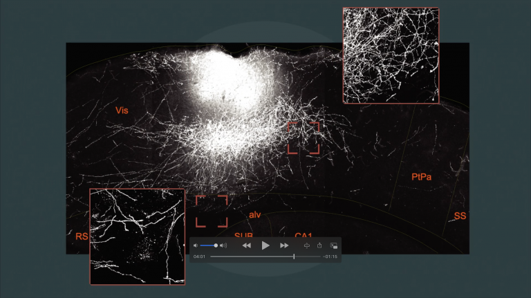
The extent to which the motor system influences the Visual system becomes apparent when the lights go out. Mark Hübener, together with colleagues, demonstrated that motor signals appear in the Visual cortex of a mouse’s brain during movement – even when the scientists had turned off the lights or when both retinas of the test animals were defective.
Neuroscientist Georg Keller from the Friedrich Miescher Institute in Basel, together with colleagues, discovered that motor signals travel from the Anterior cingulate cortex and the secondary motor Cortex to the visual cortex. Both areas are involved in controlling movement. “These signals could be a neural correlate of the efferent copy,” speculates Mark Hübener. In this way, the visual cortex is supplied with information about how much the visual world is likely to change as a result of the mouse’s movements.
“Another possibility is that expectations are derived from the experiences we have throughout our lives.” In that case, the visual system would not need a direct copy of the motor commands to generate expectations. Hübener gives an example: “We’ve already experienced millions of times how much the visual world shifts with a certain Eye movement. So we don’t need to generate an efferent copy from scratch every single time.” But of course, this learned experience must also be transmitted to the visual cortex via action potentials.
Visual system
The visual system is the part of the nervous system that processes visual information. It primarily comprises the eye, the optic nerve, the optic chiasm, the optic tract, the lateral geniculate nucleus, the optic radiation, the primary visual cortex, and the visual association cortices.
Visual cortex
The visual cortex refers to the areas of the occipital lobe that are involved in processing visual information. These include the primary visual cortex and the associative visual cortices V1 to V5. According to Brodmann, the visual cortex comprises areas 17, 18, and 19.
Anterior cingulate cortex
Anterior cingulate cortex/Anterior cingulate cortex/anterior cingulate cortex
Like the entire cingulate cortex, the anterior region of the limbic system regulates drive-controlled behavior. In the perception of pain, it is particularly associated with the affective pain component - including social pain as experienced through exclusion.
Cortex
cortex cerebri
Cortex refers to a collection of neurons, typically in the form of a thin surface. However, it usually refers to the cerebral cortex, the outermost layer of the cerebrum. It is 2.5 mm to 5 mm thick and rich in nerve cells. The cerebral cortex is heavily folded, comparable to a handkerchief in a cup. This creates numerous convolutions (gyri), fissures (fissurae), and sulci. Unfolded, the surface area of the cortex is approximately 1,800cm².
Eye
bulbus oculi
The eye is the sensory organ responsible for perceiving light stimuli – electromagnetic radiation within a specific frequency range. The light visible to humans lies in the range between 380 and 780 nanometers.
Influence at all levels?
Yet the motor system apparently already has a say at a processing stage earlier than the Visual cortex At least that is what the research by Laura Busse, a neuroscientist at LMU in Munich, suggests. She measured the electrical activity of neurons in the Lateral geniculate nucleus (LGN) of the thalamus – a part of the Visual pathway – in mice. “On average, the cells in the LGN responded more strongly when the mice were moving,” says Busse. “In addition, the cells’ ‘volume control’ – much like on a radio – was turned up higher, so that the neurons could react with greater sensitivity.”
One possible explanation: motor activity influences sensory activity even at the level of the thalamus. Laura Busse, however, remains cautious. Another explanation is also conceivable. When a person moves, they are generally more activated, leading to increased release of neurotransmitters such as acetylcholine, which influence the activity of nerve cells. “So it could be that it is not the motor influence that is responsible for the stronger response of the neurons in the LGN, but rather the generally higher state of arousal – or a combination of both.”
Mark Hübener, on the other hand, goes even a step further. There are even tentative indications that, as early as the level of the retina, the activity of neurons is influenced by movement. For Hübener, all these findings point to one conclusion: “The comparison of expected and actual sensory input does not take place at just one location in the brain, but at all possible levels of processing.” This comparison is important for the brain. After all, if the world changes in a predictable way due to one’s own movements, that is largely irrelevant. “If, on the other hand, there are more significant changes in the world – for example, because another animal is moving toward me and attacking me – then the brain and the organism must react.”
Visual cortex
The visual cortex refers to the areas of the occipital lobe that are involved in processing visual information. These include the primary visual cortex and the associative visual cortices V1 to V5. According to Brodmann, the visual cortex comprises areas 17, 18, and 19.
lateral
A positional term – lateral means "towards the side." In relation to the nervous system, it refers to a direction at right angles to the neural axis, i.e., to the right or left.
Lateral geniculate nucleus
corpus geniculatum laterale
The lateral geniculate nucleus is the section of the thalamus (the largest part of the diencephalon) where around 90% of the optic nerve axons terminate. It has a characteristic stratification into six cell layers. The nerve cells of the lateral geniculate nucleus send their projections to the visual cortex. Together with the medial geniculate nucleus, it forms the metathalamus.
Visual pathway
The visual pathway refers to the network of nerve cells involved in visual perception. In mammals, it runs from the retinal ganglion cells in the eye – as the optic nerve to the optic chiasm, then as the visual tract – via the only switching point in the lateral geniculate nucleus to the primary visual cortex.
Further reading
- Leinweber, M. et al.: A Sensorimotor Circuit in Mouse Cortex for Visual Flow Predictions. Neuron 2017 Dec 6;96(5):1204.
- Straka, H. et al.: A New Perspective on Predictive Motor Signaling. Curr Biol, 2018 Mar 5;28(5):R232-R243