The Secret to Success: Communication
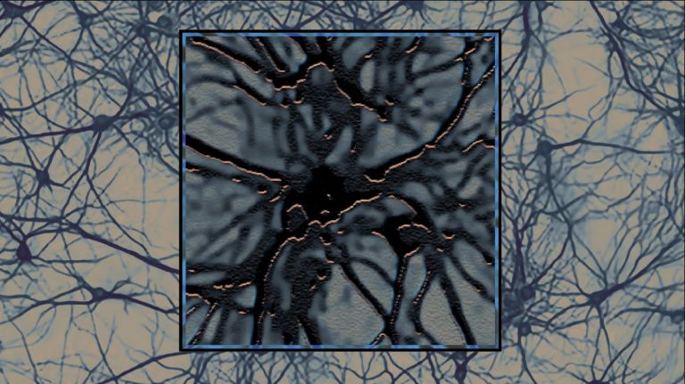
Thousands of antennae, just as many transmitters, and a super-fast data cable allow neurons to exchange information simultaneously with many other cells. In between, the signals are processed with spatial and temporal precision.
Scientific support: Prof. Dr. Jochen F. Staiger
Published: 01.12.2020
Difficulty: easy
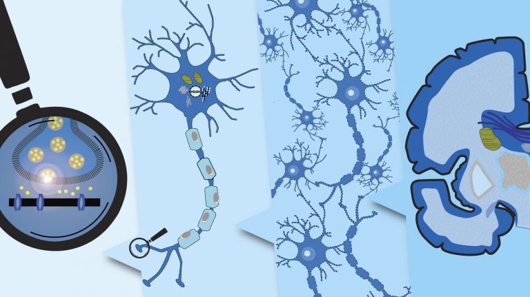
- The individual Neuron is highly specialized for communication, both structurally and functionally. It bears thousands of sensitive “antennas” around its cell body – the dendrites. It transmits electrical signals via a highly efficient “data cable” – the Axon. And it relays its messages to other cells via specialized “transmitter buttons.”
- The Synapse is the hub of neural communication. In a typical chemical synapse, the presynaptic terminal of an axon releases neurotransmitters, which diffuse across the synaptic cleft to the postsynaptic Dendrite of the partner cell and bind to receptors there.
- The postsynaptic receptors respond by opening channels that allow electrically charged particles (ions) to flow out of or into the cell. This changes the voltage of the postsynaptic cell membrane, creating a postsynaptic potential that can be excitatory or inhibitory.
- If the sum of the voltage changes across the synapses reaches a certain threshold, the postsynaptic neuron triggers an Action potential – an electrical signal that is transmitted along the axon.
- Signal transmission is more effective and faster when the axon is surrounded by an insulating Myelin sheath. This sheath is formed by specialized Glial cells called oligodendrocytes.
- At the end of the axon, the action potential triggers the release of neurotransmitters at the terminal buttons. The next chapter of communication begins.
Neuron
A neuron is a specialized cell in the nervous system that is responsible for processing and transmitting information. It receives signals via its dendrites and transmits them via its axon. Transmission occurs electrically within the neuron and, between neurons, usually chemically via synapses.
Axon
axon
The axon is the extension of the nerve cell that is responsible for conducting nerve impulses to the next cell. An axon can branch out many times, reaching a large number of downstream nerve cells. It can be more than a meter long. The axon ends in one or more synapses.
Synapse
A synapse is a connection between two neurons and serves as a means of communication between them. It consists of a presynaptic region – the terminal button of the sender neuron – and a postsynaptic region – the region of the receiver neuron with its receptors. Between them lies the synaptic cleft.
Dendrite
Tree-like branching area of nerve cells whose extensions act as a kind of antenna for receiving electrical impulses from other cells.
excitatory
Exciting synapses are described as excitatory when they depolarize the subsequent cell membrane and can thus lead to the formation of an action potential. An excitatory effect is usually produced by an exciting transmitter (messenger substance), such as glutamate. The opposite is an inhibitory synapse.
Action potential
In excitable cells (e.g., neurons or muscle cells), very rapid changes in electrical potential occur across the cell membrane. This event is the basis for signal conduction along the axon of the nerve cell. The action potential continues along the cell membrane and, according to the all-or-nothing principle, only occurs when the cell has been sufficiently excited.
Myelin
Myelin is a fatty substance produced by glial cells. It envelops the axons (long, fiber-like extensions) of nerve cells and insulates them, preventing messages from passing uncontrollably to neighboring nerve cells. This also greatly accelerates conduction velocity.
Glial cells
Glia cells are the second largest group of cells in the brain after neurons. For a long time, they were considered inactive elements of the brain, referred to as "nerve cement." Today, we know that the different types of glia cells (astrocytes, oligodendrocytes, and microglia in the CNS; Schwann cells in the PNS) perform clearly defined tasks in the nervous system. For example, they respond to pathogens, play an important role in nourishing nerve cells, and insulate nerve fibers. They account for slightly more than 50 percent of the brain's cells, compared to neurons.
Oligodendrocytes
Cells of the central nervous system that form the myelin sheath around nerve cells, thereby increasing their conduction velocity. They belong to the glial cells.
To understand the role of individual neurons in a complex system, the auditory system offers ideal conditions, according to Benedikt Grothe of Ludwig Maximilian University of Munich: “In order for us to distinguish between different sound sources occurring simultaneously, evolution has pushed neural circuits to the very limits of what is possible. There we find the entire universe of possible variables: whether a signal has an excitatory or inhibitory effect, which neurotransmitters play a role, and how the cell integrates the information.”
Thanks to multiple specializations, the neurons involved can detect the subtlest differences in the temporal and spatial patterns of sounds. Even their cell membranes are structured in such a way that only very precise signals trigger a response. This sensitivity is supported and refined by the finely tuned temporal exchange with other cells. In electrophysiological studies with desert gerbils, Grothe and his colleagues demonstrated that inhibitory signals can not only reduce but also increase the probability that the postsynaptic cell will fire.
The inhibitory signals initially lower the postsynaptic Membrane potential This briefly lowers the threshold for an action potential, making the postsynaptic Neuron more receptive to excitatory signals. Within this time window of a few microseconds, it then responds even to stimuli that would otherwise not trigger an Action potential Such precision at the single-cell level helps the brain, for example, to detect and analyze fine details of moving, faint sound sources.
The precise temporal control of inhibitory signals and the response of individual cells to them can also play a role in distinguishing between very similar sounds. The syllables “ba” and “pa,” for example, differ only by a subtle gap – or the absence thereof – at the end of the respective consonant. Some neurons involved in signal processing respond to this, among other things, by abruptly terminating an inhibitory signal. As a result, the postsynaptic neuron – which had been inhibited until then – transitions into an action potential, fires, and thereby signals the gap.
excitatory
Exciting synapses are described as excitatory when they depolarize the subsequent cell membrane and can thus lead to the formation of an action potential. An excitatory effect is usually produced by an exciting transmitter (messenger substance), such as glutamate. The opposite is an inhibitory synapse.
Membrane potential
The membrane potential is a voltage measured between the inside and outside of the cell membrane. It arises from the different distribution of electrically charged particles inside and outside the cell.
Neuron
A neuron is a specialized cell in the nervous system that is responsible for processing and transmitting information. It receives signals via its dendrites and transmits them via its axon. Transmission occurs electrically within the neuron and, between neurons, usually chemically via synapses.
Action potential
In excitable cells (e.g., neurons or muscle cells), very rapid changes in electrical potential occur across the cell membrane. This event is the basis for signal conduction along the axon of the nerve cell. The action potential continues along the cell membrane and, according to the all-or-nothing principle, only occurs when the cell has been sufficiently excited.
At the heart of neural circuits are individual nerve cells. They are the brain’s smallest unit and yet much more than mere building blocks ▸ Neurons: Building Blocks of Thinking (video). Even their external shape hints at their complexity ▸ The Neuron: Form and Function (interactive). Neurons are extremely polarized, more so than almost any other cell type. With their three iconic components, they are vaguely reminiscent of a tree: a densely branched crown of dendrites surrounding the cell body, a central trunk – the Axon – and numerous further branches in the root system of the synapses. A closer look reveals that the function and interaction of these components are what give the Neuron its special abilities – as a data octopus and a supercomputer. Neurons are specialized in gathering, processing, and distributing information. They are masters of communication. For both the exchange and the processing of information, the “outer qualities” of the nerve cell – namely, subtle differences in chemistry and electricity at the cell surface – play a crucial role.
An astonishing number of communication tools are therefore located directly in the cell membrane. It is much more than a shell that separates individual neurons from their environment. Rather, it houses channels, pumps, and receptors – the tools the nerve cell uses to receive, transmit, and process information.
Axon
axon
The axon is the extension of the nerve cell that is responsible for conducting nerve impulses to the next cell. An axon can branch out many times, reaching a large number of downstream nerve cells. It can be more than a meter long. The axon ends in one or more synapses.
Neuron
A neuron is a specialized cell in the nervous system that is responsible for processing and transmitting information. It receives signals via its dendrites and transmits them via its axon. Transmission occurs electrically within the neuron and, between neurons, usually chemically via synapses.
Communication contact points
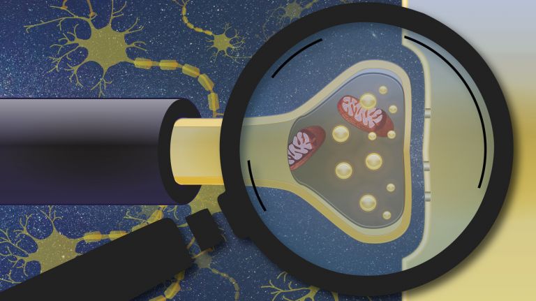
The interface for the exchange of information is the Synapse ▸ What Connects Nerves. Nerve cells are involved in up to ten thousand of these contact structures. The basic idea is simple: there is a presynaptic part that sends out signals, a synaptic cleft that the signals cross, and a postsynaptic part that receives them. The presynaptic part consists of the terminal buttons of the axons at the root region of a nerve cell, while the postsynaptic part consists of the dendrites and the cell body. Neurons thus interconnect end-to-end and usually make a strict distinction between information intake (via the dendrites and cell body) and information output (via the axon).
In a typical synapse, the Terminal button releases chemical signals (neurotransmitters) that bind to specialized Receptor proteins in the opposing postsynaptic membrane, triggering a reaction there. In addition, there are the less common electrical synapses. Here, the presynaptic and postsynaptic cells are linked via jointly formed ion channels, technically known as gap junctions. Through these, they exchange electrically charged particles (ions).
Synapse
A synapse is a connection between two neurons and serves as a means of communication between them. It consists of a presynaptic region – the terminal button of the sender neuron – and a postsynaptic region – the region of the receiver neuron with its receptors. Between them lies the synaptic cleft.
Axon
axon
The axon is the extension of the nerve cell that is responsible for conducting nerve impulses to the next cell. An axon can branch out many times, reaching a large number of downstream nerve cells. It can be more than a meter long. The axon ends in one or more synapses.
Terminal button
The end of an axon (long, fiber-like extension) of a nerve cell is called a terminal bouton if it is the presynaptic part of a synapse. The terminal bouton is a spherical thickening containing vesicles (small sacs) that store neurotransmitters (chemical messengers in the brain).
Receptor
A receptor is a protein, usually located in the cell membrane or inside the cell, that recognizes a specific external signal (e.g., a neurotransmitter, hormone, or other ligand) and causes the cell to trigger a defined response. Depending on the type of receptor, this response can be excitatory, inhibitory, or modulatory.
Electrical signals become messages
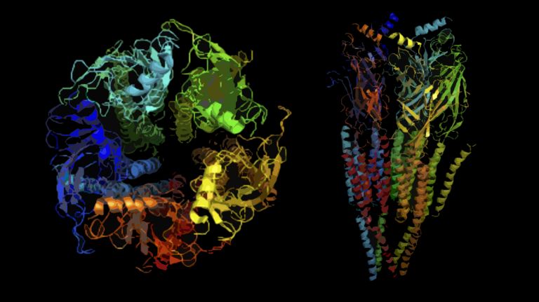
The primary language of nerve cells is electricity. The recipient cell also immediately converts the information received at a chemical Synapse back into bioelectric signals and transports these via its Axon to its own presynaptic regions. This conversion is carried out by ion channels in the cell membrane ▸ Communication Gaps: Ion Channels, which are directly or indirectly coupled to the Neurotransmitter receptors. When a transmitter molecule binds to the receptor, the Ion channel opens (or closes) and – depending on the configuration of the transmitters and channels – allows more (or fewer) positively charged sodium, calcium, or potassium ions, or negatively charged chloride ions, to cross the cell membrane. This alters the electrical charges inside the cell and in the surrounding fluid – and thus also the electrical potential between these two regions: the Membrane potential The resulting change in potential is called the postsynaptic potential.
Depending on which neurotransmitters and which channels are involved at a synapse and how the ions are distributed within the specific neuron, a postsynaptic potential can be excitatory or inhibitory and vary in strength. With its unique qualitative and quantitative signature, each postsynaptic potential functions as a piece of information in the bioelectrical communication of nerve cells.
Since a typical Neuron receives signals from other cells at many thousands of sites on its dendrites, it must integrate this abundance of signals – with their temporal and spatial patterns – into a meaningful overall message – an extremely complex computational task. The result of this computational process influences how the neuron behaves in the next step: Will it send an Action potential to its own presynaptic regions – and thus signals to other cells – or not?
At rest, a negative membrane potential of approximately -70 mV prevails, at which there are fewer positively charged particles inside the cell than outside. If the membrane potential rises above a certain threshold (approximately -55 mV) as a result of the overall assessment of the received signals, additional voltage-gated ion channels open, allowing a sudden influx of positively charged sodium ions into the cell interior. This causes the membrane potential in this region to rise even further. An action potential is generated, which then propagates in a chain reaction along the axon until it reaches the presynaptic terminals and, via the synapses located there, transmits the signal to the next neurons in the neural network. The neuron has “fired.”
Synapse
A synapse is a connection between two neurons and serves as a means of communication between them. It consists of a presynaptic region – the terminal button of the sender neuron – and a postsynaptic region – the region of the receiver neuron with its receptors. Between them lies the synaptic cleft.
Axon
axon
The axon is the extension of the nerve cell that is responsible for conducting nerve impulses to the next cell. An axon can branch out many times, reaching a large number of downstream nerve cells. It can be more than a meter long. The axon ends in one or more synapses.
Neurotransmitter
A neurotransmitter is a chemical messenger, an intermediary substance. It is released by the sender neuron at the sites of cell-cell communication and has an excitatory or inhibitory effect on the receiver neuron.
Ion channel
Ion channels are embedded in the cell membrane of nerve cells and all other cells in the body. They enable electrically charged particles, known as ions, to pass through the cell membrane into and out of the cell. They can therefore influence the membrane potential of a cell and trigger an action potential. A large number of different ion channels are known. Normally, ion channels have a specific permeability for only one type of ion, e.g., sodium ions or potassium ions. These are referred to as sodium channels or potassium channels, respectively.
Membrane potential
The membrane potential is a voltage measured between the inside and outside of the cell membrane. It arises from the different distribution of electrically charged particles inside and outside the cell.
excitatory
Exciting synapses are described as excitatory when they depolarize the subsequent cell membrane and can thus lead to the formation of an action potential. An excitatory effect is usually produced by an exciting transmitter (messenger substance), such as glutamate. The opposite is an inhibitory synapse.
Neuron
A neuron is a specialized cell in the nervous system that is responsible for processing and transmitting information. It receives signals via its dendrites and transmits them via its axon. Transmission occurs electrically within the neuron and, between neurons, usually chemically via synapses.
Action potential
In excitable cells (e.g., neurons or muscle cells), very rapid changes in electrical potential occur across the cell membrane. This event is the basis for signal conduction along the axon of the nerve cell. The action potential continues along the cell membrane and, according to the all-or-nothing principle, only occurs when the cell has been sufficiently excited.





Hop or top: the action potential
Unlike postsynaptic potentials, action potentials in vertebrates cannot be stronger or weaker. Instead, their informational content lies in whether they occur at all – and how often. The speed at which they reach their targets downstream in the information cascade is also crucial. Here, the finest differences matter – for example, a few microseconds when distinguishing between different tones in the auditory system (see box).
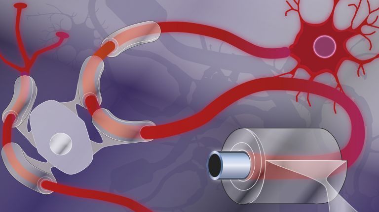
Action potentials sometimes have to travel enormous distances – axons can grow up to many meters long, depending on the organism. How fast the signals travel depends on the thickness of the axon, or rather, on how well it is electrically insulated ▸ Myelin: High Speed. In vertebrates, specialized Glial cells called Oligodendrocytes wrap around the Axon and form a Myelin sheath, comparable to the insulation around an electrical cable. Only at occasional gaps – known as Nodes of Ranvier – is the axon’s cell membrane exposed. And only here does the sudden ion exchange and the associated change in electrical voltage, which the Action potential needs to continue its journey, occur effectively. Within the electrically insulated segments of the axon, however, the voltage difference is transmitted almost instantaneously by an electric field. This field also propels ions further away within the cell sufficiently to create the conditions at the next node of Ranvier for the voltage-gated ion channels there to open. The action potential thus “jumps” from node to node.
When the action potential finally reaches the axon terminals, it triggers the opening of additional ion channels there, allowing calcium ions to flow into the cell. The increased concentration of calcium ions, in turn, serves as a signal for the release of neurotransmitters into the synaptic cleft, and thus the signal reaches the next cell. This concludes the neuron’s current contribution to communication. However, the Neuron can also respond to the communication in which it participates over the long term – for example, by strengthening frequently used synapses, weakening or breaking down rarely used ones, or forming new synapses. Through this synaptic plasticity, the communicative capacities of these neurons and the brain are constantly changing – we learn, form new memories, and forget others.
Glial cells
Glia cells are the second largest group of cells in the brain after neurons. For a long time, they were considered inactive elements of the brain, referred to as "nerve cement." Today, we know that the different types of glia cells (astrocytes, oligodendrocytes, and microglia in the CNS; Schwann cells in the PNS) perform clearly defined tasks in the nervous system. For example, they respond to pathogens, play an important role in nourishing nerve cells, and insulate nerve fibers. They account for slightly more than 50 percent of the brain's cells, compared to neurons.
Oligodendrocytes
Cells of the central nervous system that form the myelin sheath around nerve cells, thereby increasing their conduction velocity. They belong to the glial cells.
Axon
axon
The axon is the extension of the nerve cell that is responsible for conducting nerve impulses to the next cell. An axon can branch out many times, reaching a large number of downstream nerve cells. It can be more than a meter long. The axon ends in one or more synapses.
Myelin
Myelin is a fatty substance produced by glial cells. It envelops the axons (long, fiber-like extensions) of nerve cells and insulates them, preventing messages from passing uncontrollably to neighboring nerve cells. This also greatly accelerates conduction velocity.
Action potential
In excitable cells (e.g., neurons or muscle cells), very rapid changes in electrical potential occur across the cell membrane. This event is the basis for signal conduction along the axon of the nerve cell. The action potential continues along the cell membrane and, according to the all-or-nothing principle, only occurs when the cell has been sufficiently excited.
Neuron
A neuron is a specialized cell in the nervous system that is responsible for processing and transmitting information. It receives signals via its dendrites and transmits them via its axon. Transmission occurs electrically within the neuron and, between neurons, usually chemically via synapses.
Just one cell among billions?
Given that there are approximately 86 billion neurons in the human brain, the question arises as to what role a single cell can play in this complex and dynamic structure. Considering that each Neuron processes many thousands of temporally and spatially structured inhibitory and excitatory signals per millisecond, translates them into electrical signal frequencies of up to 1,000 hertz, and finally transmits the message via precisely tuned axons, the answer is: a considerable one. The supercomputer “SpiNNaker,” which went into operation in 2018 and attempts to simulate one billion interconnected nerve cells, has over one million processing cores, each of which is designed to model 100 neurons with 10,000 synapses. A current high-performance laptop has 8 cores. If we do a rough calculation, an average nerve cell in the human brain still manages to achieve one-eight-hundredth of the computing power of a MacBook Pro. Well, that was the situation in 2020. Still: Not bad for just a single cell.
Neuron
A neuron is a specialized cell in the nervous system that is responsible for processing and transmitting information. It receives signals via its dendrites and transmits them via its axon. Transmission occurs electrically within the neuron and, between neurons, usually chemically via synapses.
excitatory
Exciting synapses are described as excitatory when they depolarize the subsequent cell membrane and can thus lead to the formation of an action potential. An excitatory effect is usually produced by an exciting transmitter (messenger substance), such as glutamate. The opposite is an inhibitory synapse.
Further reading
- Hammond, Constance: Cellular and Molecular Neurophysiology, Academic Press (2015)
- Koch, Christof: Biophysics of Computation: Information Processing in Single Neurons, Oxford University Press: New York (1999)
- Beiderbeck B et al: Precisely timed inhibition facilitates action potential firing for spatial coding in the auditory brainstem. Nature Communications. (2018)9:1771 ( zum Volltext ).