Connecting Nerves
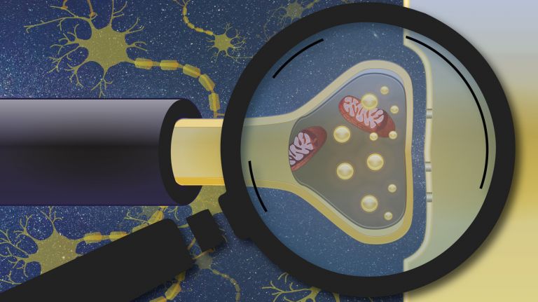
It’s all about the right connections: Synapses enable us to respond flexibly to situations and to learn.
Scientific support: Prof. Dr. Jochen F. Staiger
Published: 01.12.2020
Difficulty: easy
- The connections between nerve cells via synapses allow us to adapt our behavior flexibly to different situations. Neurons that produce Dopamine influence sensory Perception and, consequently, decision-making.
- Some synaptic activity alters the response of downstream neurons only briefly, thereby influencing an immediate decision. Recurring activity can lead to long-term changes in communication between neurons. This is how we learn.
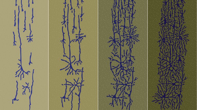
- During learning, extensions a few thousandths of a millimeter long grow on nerve cells.
- Learning at the synaptic level can be studied using two-photon microscopy
- Computer models also help researchers better understand the intricate mechanisms at synapses
Dopamine
Dopamine is an important neurotransmitter in the central nervous system that belongs to the catecholamine group. It plays a role in motor function, motivation, emotion, and cognitive processes. Disruptions in the function of this transmitter play a role in many brain disorders, such as schizophrenia, depression, Parkinson's disease, and substance dependence.
Perception
The term describes the complex process of gathering and processing information from stimuli in the environment and from the internal states of a living being. The brain combines the information, which is perceived partly consciously and partly unconsciously, into a subjectively meaningful overall impression. If the data it receives from the sensory organs is insufficient for this, it supplements it with empirical values. This can lead to misinterpretations and explains why we succumb to optical illusions or fall for magic tricks.
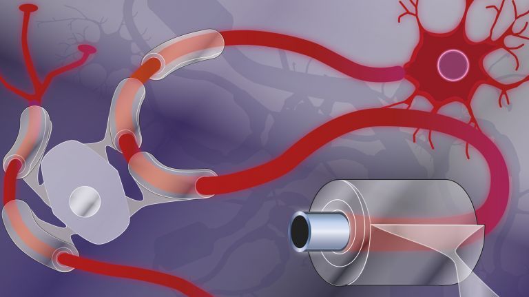
To keep our gaze stable even as we move, the nervous system has to employ some clever tricks. The Vestibular system in the inner Ear detects head movement. It then sends signals to the Eye muscles, causing the eyes to move and thus compensate for the head movement. For example, if you move your head to the right, your eyes move to the left. However, neurobiologist Hans Straka (1961–2022) of Ludwig Maximilian University of Munich was able to show that, at least for very rhythmic and uniform movements, the signals come directly from the Spinal cord Straka observed a particular flexibility in tadpoles. While a tadpole swims in a back-and-forth motion to the right and left, the adult frog moves forward in jerky bursts using its legs. This requires entirely different eye movements to keep the gaze stable. And these changed requirements are accommodated by the nervous system. The basis for this flexibility is a change in the neural connections between the spinal cord and the control centers for the eye muscles in the brainstem. “This synaptic Plasticity ensures that functionally correct eye movements are triggered regardless of the movement pattern,” says Straka.
Vestibular system
Vestibular apparatus/Organon vestibulare/vestibular organ
The vestibular system is part of the inner ear. Its sensors are located in the semicircular canals. As part of the balance system, it detects circular movements (rotations), acceleration, and gravity.
Ear
auris
The ear is not only the organ of hearing, but also of balance. A distinction is made between the outer ear with the auricle and external auditory canal, the middle ear with the eardrum and ossicles, and the actual hearing and balance organ, the inner ear with the cochlea and semicircular canals.
Eye
bulbus oculi
The eye is the sensory organ responsible for perceiving light stimuli – electromagnetic radiation within a specific frequency range. The light visible to humans lies in the range between 380 and 780 nanometers.
Spinal cord
medulla spinalis
The spinal cord is the part of the central nervous system located in the spine. It contains both the white matter of the nerve fibers and the gray matter of the cell nuclei. Simple reflexes such as the knee-jerk reflex are already processed here, as sensory and motor neurons are directly connected. The spinal cord is divided into the cervical, thoracic, lumbar, and sacral spinal cord.
Plasticity
Neuroplasticity
The term neuroplasticity describes the ability of synapses, nerve cells, and entire areas of the brain to change structurally and functionally depending on the degree to which they are used. Synaptic plasticity refers to the adaptation of the signal transmission strength of synapses to the frequency and intensity of incoming stimuli, for example in the form of long-term potentiation or depression. In addition, the size, interconnection, and activity patterns of different areas of the brain also change depending on their use. This phenomenon is referred to as cortical plasticity when it specifically affects the cortex.
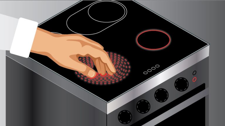
If we were mere stimulus-response machines, our lives would be quite boring, predictable, and devoid of purpose. Imagine, for example, if we always pounced on food the moment we saw it – even when we weren’t hungry at all. Fortunately, however, we are generally quite flexible in our behavior. This is made possible by the interconnection of nerve cells via synapses. ▸ Synapses: Interfaces of Life
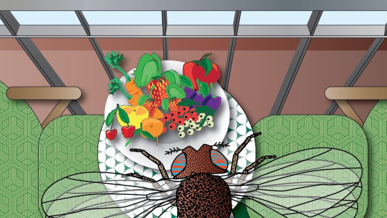
Ilona Grunwald Kadow has been closely examining how the connections between our brain cells affect our behavior for years. “Our decisions are based on how we assess situations,” says the neuroscientist, formerly at the Technical University of Munich and now at the University Hospital Bonn. “This assessment, in turn, is determined by our experience, our current needs, and what our sensory Perception is presenting to us at that moment.” For example, it makes a big difference whether an organism is hungry or not. Hungry animals place a higher value on smells and tastes than usual. This is because, in such cases, the “reward hormone” Dopamine is released in greater quantities at the synapses in various regions of the brain. “Neurons that produce dopamine influence sensory perception and thus allow behavior to be flexibly adapted to current situations,” says Grunwald Kadow.
In a study published 2020, she examined the response of all dopamine-producing neurons in over 400 living fruit flies when exposed to Taste and smell stimuli. The result: Certain neurons appear, for example, to detect whether the fly is currently moving, resting, or grooming itself. If the fly smells something enticing and the pleasant scent intensifies when the fly moves, the signal is: Keep moving in this direction. Grunwald is not studying the cell bodies, but rather the synapses. Upstream neurons send information – such as odors – to downstream neurons, which then control whether the organism moves or not.
Kadow has already shed light on what such communication might look like in a study published in 2015. Together with colleagues, she exposed fruit flies to two odors that trigger conflicting “emotions” in the animals: the enticing food odor of vinegar, and CO₂ which can signal danger. When the flies detect CO₂, an innate flight response is triggered. However, this does not exactly make decision-making any easier for the flies: CO₂ is also produced – in a rather tricky twist – by overripe fruit, a coveted food source for many insects. In such cases, foraging flies must therefore be able to ignore their innate aversion to CO₂. The study revealed that the smell of vinegar did indeed activate certain dopaminergic neurons. These, in turn, suppressed activity in synapses that respond to negative odors such as CO₂. As a result, the hungry flies were less responsive to the potential danger and were able to focus on the source of the enticing scent.
Perception
The term describes the complex process of gathering and processing information from stimuli in the environment and from the internal states of a living being. The brain combines the information, which is perceived partly consciously and partly unconsciously, into a subjectively meaningful overall impression. If the data it receives from the sensory organs is insufficient for this, it supplements it with empirical values. This can lead to misinterpretations and explains why we succumb to optical illusions or fall for magic tricks.
Dopamine
Dopamine is an important neurotransmitter in the central nervous system that belongs to the catecholamine group. It plays a role in motor function, motivation, emotion, and cognitive processes. Disruptions in the function of this transmitter play a role in many brain disorders, such as schizophrenia, depression, Parkinson's disease, and substance dependence.
Taste
The sensory impression we refer to as "taste" results from the interaction between our senses of smell and taste. In terms of sensory physiology, however, "taste" is limited to the impression conveyed to us by the taste receptors on the tongue and in the surrounding mucous membranes. It is currently assumed that there are five different types of taste receptors that specialize in the taste qualities sweet, sour, salty, bitter, and umami. In 2005, scientists also identified possible taste receptors for fat, whose role as a distinct taste quality is still being investigated.
From firing to wiring
“Some activity of dopaminergic synapses alters the response of downstream neurons only briefly, thereby influencing an immediate decision,” says Kadow. “But above all, recurring activity can lead to long-term changes in communication between neurons.” This is how living organisms learn. During learning, tiny extensions form on nerve cells. To establish a synaptic connection, these extensions on opposing nerve cells grow into mushroom-like structures. On the upstream side of the Neuron – where the stimulus originates – axonal terminals form. On the opposite, receiving side, so-called Dendritic spines develop. The interaction between these two projections enables the exchange of information between different nerve cells. The rule of learning is: Neurons that fire together wire themselves together. ▸ The Nervous System – A Master of Adaptation.
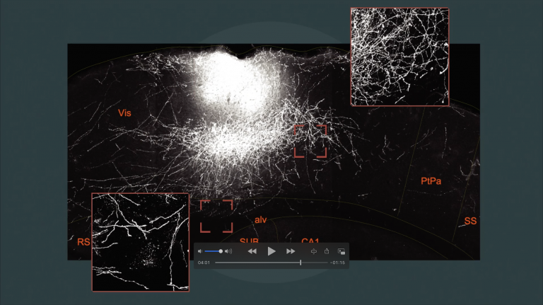
To observe new connections forming in real time, Mark Hübener relies on two-photon microscopy. “It allows us to examine even very fine structures in the intact nervous system,” says the neurobiologist at the Max Planck Institute for Biological Intelligence in Martinsried, near Munich. “The advantage is that you can look deeper into the tissue than with a normal microscope – up to one millimeter.” This is because two-photon microscopy uses light with a long wavelength – namely, infrared light. It penetrates tissue more easily than shorter-wavelength light. Furthermore, it is much less harmful to the tissue than, for example, UV light.
Neuron
A neuron is a specialized cell in the nervous system that is responsible for processing and transmitting information. It receives signals via its dendrites and transmits them via its axon. Transmission occurs electrically within the neuron and, between neurons, usually chemically via synapses.
Dendritic spines
Mushroom- or button-shaped protrusions on dendrites, at the tip of which there is usually a synapse, which is the site of communication between two nerve cells. The spines increase the surface area of dendrites, which thus have space for additional synapses. They play an important role in synaptic plasticity. The dendritic spines can swell and shrink depending on activation.
Adaptation
Adaptation refers to the process by which the sensory organs, the perceptual system, or the entire organism adjusts to the intensity and quality of stimuli and to changes in environmental conditions. In visual adaptation, for example, the pupil and the sensitivity of the photoreceptors regulate themselves according to the prevailing light conditions.
Intelligence
Intelligence
Collective term for human cognitive performance. According to British psychologist Charles Spearman, cognitive performance in different areas correlates with a general factor (g factor) of intelligence. This means that intelligence can be expressed as a single value. American psychologist Howard Gardner, among others, has developed a counter-concept to this, known as the "theory of multiple intelligences." According to this theory, intelligence develops independently in the following eight areas: linguistic, logical-mathematical, musical-rhythmic, visual-spatial, bodily-kinesthetic, naturalistic, intrapersonal, and interpersonal.
Learning: a spiny path
“This method is particularly well-suited for our research into synaptic Plasticity because synapses are very small structures and are often located deep within the brain.” Both axonal terminals and Dendritic spines are only two-to-three-thousandths of a millimeter in size. In his experiments, Hübener, for example, exposes mice to a new experience. Over the course of days or weeks, he can then repeatedly check whether this is accompanied by the formation of dendritic spines. “If, for example, a dendritic spine on the downstream side of the Synapse grows larger or forms for the first time, then we know that synaptic connections have become stronger or have formed anew.” A change has thus taken place that underlies learning. In the same way, two-photon microscopy can be used to examine changes in the axonal terminals on the presynaptic side of the synapse.
Plasticity
Neuroplasticity
The term neuroplasticity describes the ability of synapses, nerve cells, and entire areas of the brain to change structurally and functionally depending on the degree to which they are used. Synaptic plasticity refers to the adaptation of the signal transmission strength of synapses to the frequency and intensity of incoming stimuli, for example in the form of long-term potentiation or depression. In addition, the size, interconnection, and activity patterns of different areas of the brain also change depending on their use. This phenomenon is referred to as cortical plasticity when it specifically affects the cortex.
Dendritic spines
Mushroom- or button-shaped protrusions on dendrites, at the tip of which there is usually a synapse, which is the site of communication between two nerve cells. The spines increase the surface area of dendrites, which thus have space for additional synapses. They play an important role in synaptic plasticity. The dendritic spines can swell and shrink depending on activation.
Synapse
A synapse is a connection between two neurons and serves as a means of communication between them. It consists of a presynaptic region – the terminal button of the sender neuron – and a postsynaptic region – the region of the receiver neuron with its receptors. Between them lies the synaptic cleft.

The right place
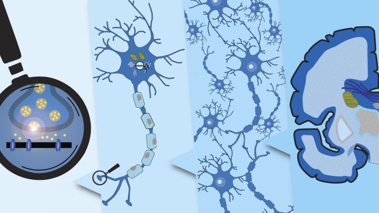
When a Synapse is learning and needs to be restructured for that purpose, various molecular processes come into play. In nerve cells, a portion of the RNA – the transcript of DNA – is translated into new proteins not only in the cell body but also locally at the dendrites. To achieve this, the transcript must be transported specifically to the synapses. However, how the cell ensures that the correct blueprint is delivered to the right address has remained largely unknown until now. Using fluorescence microscopy, Michael Kiebler from the Biomedical Center at Ludwig Maximilian University of Munich examined cell cultures from the rat Hippocampus. As it turned out, the same RNA circulated repeatedly from the cell body to the processes and back until it was needed by a synapse. In this process, specific recognition sequences in the RNA serve as a kind of “postmark” for transport and ensure that the transcript reaches the correct locations within the cell. The transcript can then be translated into proteins for the necessary remodeling of the synapse.
Synapse
A synapse is a connection between two neurons and serves as a means of communication between them. It consists of a presynaptic region – the terminal button of the sender neuron – and a postsynaptic region – the region of the receiver neuron with its receptors. Between them lies the synaptic cleft.
Hippocampus
The hippocampus is the largest part of the archicortex and an area in the temporal lobe. It is also an important part of the limbic system. Functionally, it is involved in memory processes, but also in spatial orientation and learning. It comprises the subiculum, the dentate gyrus, and the Ammon's horn with its four fields CA1-CA4.
Changes in the structure of the hippocampus due to stress are associated with chronic pain. The hippocampus also plays an important role in the amplification of pain through anxiety.
Synapses in the computer
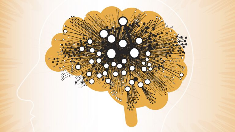
In addition to experiments, computer models also help researchers get to the bottom of the intricate mechanisms at the synapses. Christian Leibold – now at the University of Freiburg – was investigating what happens between the two ears: “Sound takes different amounts of time to reach the left and right ears,” says the neurobiologist. “The brain can localize the sound based on this time difference.” Neurons in the medial superior olive (MSO) use their sensitivity to these time differences to generate a neural map of the acoustic space. Using computer models, Leibold simulated the sound processing of MSO neurons. These nerve cells have four input pathways at the synapse. Two excitatory pathways from the right and left ears, respectively. These increase the likelihood that the Neuron will fire. In addition, there are two inhibitory pathways that have the opposite effect. In the computer model, Leibold and his colleagues then investigated how fast these excitatory and inhibitory potentials must be at the Synapse to explain the neurons’ sensitivity to time differences, as demonstrated in experiments. A similar problem arises for the nerve cells in the midbrain, which process the MSO’s activity to resolve complex sound situations – for example, multiple speakers. In their models, the researchers need only adjust a few parameters, such as changing the ratio of excitatory to inhibitory potentials. “In fact, it’s enough to tweak just a few parameters to explain all the cell’s responses observed in the experiment,” says Leibold. The researchers are like the synapses themselves: they are constantly learning.
medial
A positional term – medial means "towards the middle." In relation to the nervous system, it refers to a direction toward the body, away from the sides.
excitatory
Exciting synapses are described as excitatory when they depolarize the subsequent cell membrane and can thus lead to the formation of an action potential. An excitatory effect is usually produced by an exciting transmitter (messenger substance), such as glutamate. The opposite is an inhibitory synapse.
Neuron
A neuron is a specialized cell in the nervous system that is responsible for processing and transmitting information. It receives signals via its dendrites and transmits them via its axon. Transmission occurs electrically within the neuron and, between neurons, usually chemically via synapses.
Synapse
A synapse is a connection between two neurons and serves as a means of communication between them. It consists of a presynaptic region – the terminal button of the sender neuron – and a postsynaptic region – the region of the receiver neuron with its receptors. Between them lies the synaptic cleft.
Further reading
- Siju, K.P et al.: Valence and State-Dependent Population Coding in Dopaminergic Neurons in the Fly Mushroom Body. In: Current Biology, Volume 30 (11), P2104-2115.e4, June 08, 2020. doi: https://doi.org/10.1016/j.cub.2020.04.037
- Bauer, K.E. et al.: Live cell imaging reveals 3'-UTR dependent mRNA sorting to synapses. Nat Commun. 2019 Jul 18;10(1):3178. doi: 10.1038/s41467-019-11123-x.
First published on December 1, 2020
Last updated on May 5, 2026