Communication Gaps: Ion Channels
Ion channels hold the key to understanding the electrical activity of nerve cells. Without them, literally nothing works.
Scientific support: Prof. Dr. Helmut Kettenmann
Published: 01.12.2020
Difficulty: intermediate
- Ion channels are integral membrane proteins found in all cells and characterized by three properties: They allow ions to pass through the otherwise impermeable membrane; they do so (relatively) specifically for certain ions only; and they open and close in response to specific electrical, chemical, or mechanical signals.
- Ion channels can be divided into three groups: voltage-gated ion channels, ligand-gated ion channels, and mechanically activated ion channels.
- Many diseases can be attributed to changes in the structure of ion channels.
- Many medications, drugs, and toxins act as ligands at receptors for neurotransmitters.
A key ability of the brain is its capacity to respond – immediately and to stimuli originating both internally and externally. This can be viewed as the primary function of the nervous system: receiving signals, transmitting them, and responding to them. In some cases, surprisingly small stimuli are sufficient to trigger an electrical signal in a nerve cell. In the Photoreceptors of our retina, a single photon can be enough to do so. These almost unimaginably tiny stimuli cause a change in the electrical voltage across the membrane of the nerve cells, which relay this change as a signal over distances that are sometimes astonishing, until we can perceive it and react to it. All of this is based on the structure and function of ion channels. They hold the key to understanding the electrical activity of nerve cells and the versatility of their signals.
Photoreceptors
Photoreceptors are the light-sensitive cells of the retina; they convert light into electrical potentials. There are approximately 127 million photoreceptors in the retina, including seven million cones and 120 million rods.
What are ion channels?
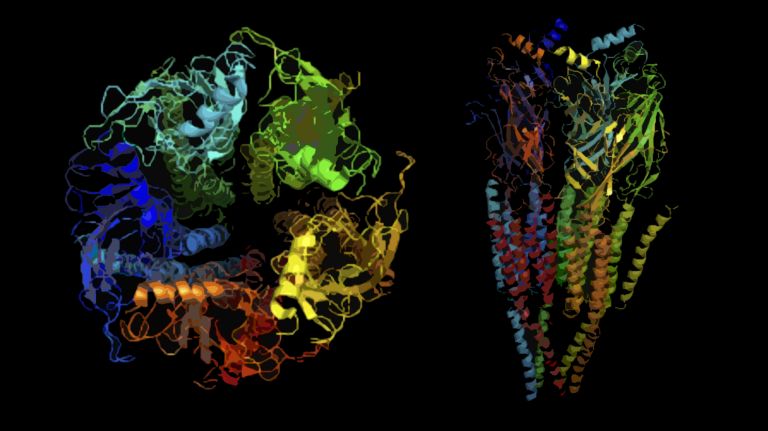
Ion channels are large proteins found in the membranes of all body cells. They span the membrane, protruding both inward and outward; they are therefore called integral membrane proteins. The ion channels in neurons do not differ in principle from those in other body cells. Depending on the type of cell in which they are found, different ion channels perform different functions. But they all share three essential properties:
- They allow ions to pass through the otherwise impermeable membrane,
- They do so (relatively) specifically for certain ions only – that is, they are selective,
- They open and close in response to specific electrical, chemical, or mechanical signals.
What makes neuronal ion channels special is their speed and high conductivity – to be precise, about 12 million ions per second. It is exactly this massive flow of electrical charges that forms the fundamental prerequisite for utilizing the mechanism of signal transmission – even though the current generated in the process amounts to just under two picoamperes, or two trillionths of an ampere. What is astonishing is how selective (though not exclusive) the channels are with regard to the ions they allow to pass through. For example, potassium channels are about a hundred times more conductive for potassium than for sodium, while sodium channels are about twenty times more conductive for sodium than for potassium. In addition to positively charged ions (cations) such as sodium and potassium, the passage of negatively charged ions (anions), such as chloride, is also regulated. However, the principle of operation is always the same.
How do ion channels work?
Functionally, the channels are complemented by pumps – usually very complex proteins that are also located in the membrane. It is these pumps that separate sodium and potassium from one another. They expend energy to continuously pump sodium and chloride out of the cell and potassium into it. And because these substances are ions – that is, electrically charged particles – their unequal distribution creates an electric charge or polarity. Thus, a voltage exists across the membrane, measured in volts. At rest, nerve cells are negatively charged by about 70 thousandths of a volt compared to their surroundings. Reducing this value – that is, depolarizing the cell – means that energy is released: the cell is excited. Conversely, increasing this value – that is, building up voltage and hyperpolarizing the cell – means that the cell is inhibited. Both the Membrane potential and the concentration differences between the ions inside and outside the cell are crucial in determining whether and how the nerve cell responds to a stimulus. To understand how it is possible that each channel allows (almost) only one type of ion to pass through, we need to take a closer look at both the ions and the channels:
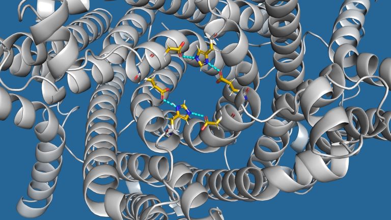
Sodium and potassium both carry a positive charge. However, because sodium is smaller, this charge is distributed over a smaller volume, resulting in a stronger electric field for sodium. Although water is electrically neutral on the surface, it still possesses a certain degree of polarity. Water molecules cluster around charged particles – this is known as the hydration shell, which is slightly larger for sodium than for potassium, due to sodium’s stronger electric field. However, the sheer size of the ions, including their hydration shells, is not the decisive factor in the selectivity of the channels. In any case, it could only explain the selectivity of the potassium channels. More relevant is the inner wall of the channel. There are amino acids located there that also carry charges. The regions with charged amino acids function as a kind of molecular sieve that selects individual ions. Because the channel pore is too small to allow an ion to pass through along with its hydrate shell, the ions lose their water coating here. For a very brief moment, the ion interacts with the amino acids, which accelerate it as it passes through the channel via electrostatic and diffusion forces and push it through the channel pore. However, this works really well only if the energy provided by the charged amino acids corresponds to the energy the ion loses by shedding its water shell. Thus, one can deduce which ion the channel protein selects based solely on its structure. This is because highly charged amino acids, such as aspartate or glutamate, interact more strongly with the larger field of sodium ions, thereby giving it more momentum and thus selecting this cation. If the inner surface of the channel contains amino acids with a lower charge, this channel tends to prefer potassium, because sodium does not have enough energy to be forced through the channel.
Membrane potential
The membrane potential is a voltage measured between the inside and outside of the cell membrane. It arises from the different distribution of electrically charged particles inside and outside the cell.


How do ion channels open and close?
Apart from that, the structure of the channel proteins is responsible for something else as well. Such a channel is not a permanently open hole in the membrane. Otherwise, the ion gradient established by the pumps would be depleted within a very short time. There are therefore opening and closing mechanisms, also referred to as “gating.” More specifically, there are three such mechanisms, allowing channels to be divided into three main classes:
- Voltage-gated ion channels,
- Ligand-gated ion channels,
- Mechanically activated ion channels.
If the inner surface of the channel is connected to the cell’s cytoskeleton, then tensile, compressive, or shear forces can open or close the pore. The result is a mechanically activated ion channel.
The voltage dependence of channels arises from the fact that the charged amino acids, firmly bound within the channel pore, cannot leave their positions but, of course, still respond to the Membrane potential The voltage shifts the charge-carrying amino acids until the channel pore becomes impassable. Only when the voltage is removed does the channel pore straighten out and the channel become passable.
However, this ingenious mechanism also gives rise to problems – at least when genes encoding ion channels are mutated at a site relevant to the opening mechanism. Even a change in a single genetic building block can cause one channel to become less conductive – or another to become excessively conductive – for certain ions. As a result, nerve cells may respond to an input signal not with a single output signal, but with a whole series of them. This mechanism underlies certain forms of epilepsy, for example. Here, the anion chloride comes into play once again. This is because the positively charged cations need an antagonist; otherwise, the activity they carry runs amok and nothing can stop it.
Inhibition and excitation form a delicate balance that can easily be disrupted by such minute changes – sometimes with dramatic consequences. This clearly demonstrates why it is so clinically relevant to study ion channels in depth. And why ligand-gated ion channels are the subject of such intensive research in this field. This is because a channel’s voltage dependence can hardly be altered once it has been developed.Ligand-gated ion channels, on the other hand, only open when a signaling molecule – such as a messenger substance or Neurotransmitter – binds to the outer surface. This, too, causes the shape of the channel protein to change slightly, and the pore opens. With this conformational change, the nerve signal begins in the postsynaptic region. Positively charged sodium ions flow into the cell, and potassium flows out. This causes the voltage to drop; the cell depolarizes until the voltage-gated sodium channels open – all at the same time. The massive influx causes the potential to overshoot far into the positive range, creating a large electromagnetic field that extends beyond the sheath cells and opens more distant voltage-gated channels. The signal thus jumps across the sheath cells, which is why this is referred to as saltatory conduction.
Membrane potential
The membrane potential is a voltage measured between the inside and outside of the cell membrane. It arises from the different distribution of electrically charged particles inside and outside the cell.
Inhibition
Neuronal inhibition describes the phenomenon whereby a sender neuron sends an impulse to a receiver neuron, causing the latter's activity to decrease. The most important inhibitory neurotransmitter is GABA.
Neurotransmitter
A neurotransmitter is a chemical messenger, an intermediary substance. It is released by the sender neuron at the sites of cell-cell communication and has an excitatory or inhibitory effect on the receiver neuron.
A starting point for toxins and drugs
Because all of this begins with a transmitter binding to the Receptor – and because this binding opens the corresponding channel through a conformational change that depends on the structure of the transmitter molecule – there is an opportunity here for regulatory intervention. One “only” needs to find a molecule that is similar enough to the natural transmitter to bind to the receptor, but different enough to cause the channel protein to adopt a different conformation. In this way – for example, through the use of certain medications – ion channels can be made to open or close for longer or shorter periods, or more quickly or more slowly. This is exactly what happens in epilepsy treatment, when excessively conductive voltage-gated channels are inhibited.
A large part of pharmacological research involves finding substances that are related to natural neurotransmitters but act slightly differently at the receptors. The next step is to determine how this affects the overall system. Nature also makes use of this mechanism. For example, many toxins found in the natural world are substances that interfere with ion channels – sometimes reversibly (such as the arrow poison curare) and sometimes permanently (such as the snake venom alpha-bungarotoxin).
Ion channels are therefore not only key to the electrical activity of nerve cells – and thus to our ability to move, to perceive or respond to environmental stimuli, to feel, and to think – but they are also crucial for understanding many diseases. Knowledge of the structure and function of ion channels can help develop specific therapies to treat neurological and mental illnesses.
Receptor
A receptor is a protein, usually located in the cell membrane or inside the cell, that recognizes a specific external signal (e.g., a neurotransmitter, hormone, or other ligand) and causes the cell to trigger a defined response. Depending on the type of receptor, this response can be excitatory, inhibitory, or modulatory.
Further reading
Ion Channells in "Principles of Neural Science" 5th Edition, Eric Kandel (Hrsg.), New York, 2013