
Myelin: High Speed
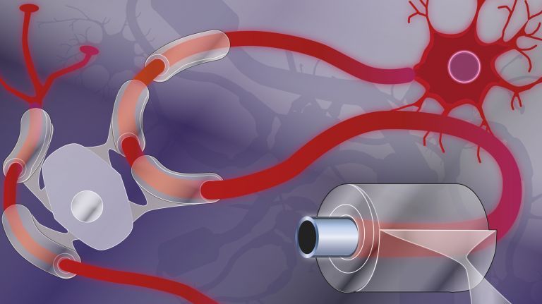
Oligodendrocytes wrap the axons of nerve cells in an insulating layer of myelin so that electrical signals can travel faster. This requires careful planning of the exact characteristics of the “road sections.”
Scientific support: Prof. Dr. Rudolf Martini
Published: 01.12.2020
Difficulty: intermediate
- Nerve cells communicate by sending action potentials on a long journey to the synapses at the end of their axons – a complex and relatively slow process.
- The Axon only becomes a “superhighway” with the help of Oligodendrocytes. These Glial cells wrap around multiple axons in sections, supply them with energy, and electrically insulate the wrapped segments with their biomembrane, known as Myelin. Within the wrapped segments, the Action potential propagates much faster.
- Between two myelin segments, the cell membrane is exposed. In these so-called Ranvier nodes, the action potential is regenerated in response to each segment and thus jumps from node to node until it reaches the end of the axon.
- How fast an action potential travels along a myelinated axon depends on many factors: the axon’s diameter, the thickness of the myelin sheath, and the length of the myelin segments. These factors vary from cell to cell as well as across different regions of an axon and change during developmental or learning processes.
- Little is yet known about how nerve cells and oligodendrocytes communicate with each other to determine myelination patterns. However, there is ample evidence that both cell types influence one another.
Axon
axon
The axon is the extension of the nerve cell that is responsible for conducting nerve impulses to the next cell. An axon can branch out many times, reaching a large number of downstream nerve cells. It can be more than a meter long. The axon ends in one or more synapses.
Oligodendrocytes
Cells of the central nervous system that form the myelin sheath around nerve cells, thereby increasing their conduction velocity. They belong to the glial cells.
Glial cells
Glia cells are the second largest group of cells in the brain after neurons. For a long time, they were considered inactive elements of the brain, referred to as "nerve cement." Today, we know that the different types of glia cells (astrocytes, oligodendrocytes, and microglia in the CNS; Schwann cells in the PNS) perform clearly defined tasks in the nervous system. For example, they respond to pathogens, play an important role in nourishing nerve cells, and insulate nerve fibers. They account for slightly more than 50 percent of the brain's cells, compared to neurons.
Myelin
Myelin is a fatty substance produced by glial cells. It envelops the axons (long, fiber-like extensions) of nerve cells and insulates them, preventing messages from passing uncontrollably to neighboring nerve cells. This also greatly accelerates conduction velocity.
Action potential
In excitable cells (e.g., neurons or muscle cells), very rapid changes in electrical potential occur across the cell membrane. This event is the basis for signal conduction along the axon of the nerve cell. The action potential continues along the cell membrane and, according to the all-or-nothing principle, only occurs when the cell has been sufficiently excited.
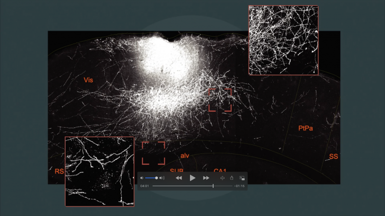
Neurons and Glial cells interact in the developing nervous system like dancers in a ballet. This can be observed particularly well in zebrafish. It offers researchers two major advantages: First, using genetic and molecular biological techniques, all kinds of processes can be manipulated and made to glow using fluorescent proteins. And second, the embryos and young fish larvae are transparent, allowing researchers to film the processes inside the larvae live and in color under a microscope.
In zebrafish, the camera is there to capture the moment when neurons and glial cells extend their tentacles, probe their surroundings and neighbors, or transmit signals. In this way, researchers observe the formation of new Myelin as Oligodendrocytes mature, as well as when previously demyelinated segments are re-myelinated.
Through a skillful combination of manipulation and observation, researchers can, in the best-case scenario, decipher the underlying mechanisms. If scientists block the release of neurotransmitters at the synapses during an experiment, they can later observe that the oligodendrocytes wrap less myelin around the axons. This leads to the conclusion that the activity of neurons helps determine how well their axons are insulated.
Glial cells
Glia cells are the second largest group of cells in the brain after neurons. For a long time, they were considered inactive elements of the brain, referred to as "nerve cement." Today, we know that the different types of glia cells (astrocytes, oligodendrocytes, and microglia in the CNS; Schwann cells in the PNS) perform clearly defined tasks in the nervous system. For example, they respond to pathogens, play an important role in nourishing nerve cells, and insulate nerve fibers. They account for slightly more than 50 percent of the brain's cells, compared to neurons.
Myelin
Myelin is a fatty substance produced by glial cells. It envelops the axons (long, fiber-like extensions) of nerve cells and insulates them, preventing messages from passing uncontrollably to neighboring nerve cells. This also greatly accelerates conduction velocity.
Oligodendrocytes
Cells of the central nervous system that form the myelin sheath around nerve cells, thereby increasing their conduction velocity. They belong to the glial cells.
Neurons, the “gray matter” in the brain, rightly deserve their reputation as masters of computation. But only once they are clad in white do they also perform communication wonders. To exchange information with many, many other neurons, nerve cells need axons. Through these “data cables,” they make contact with one another and form extensive networks. In doing so, they span distances of up to several meters – a huge distance in the cellular cosmos. To accomplish this, axons need either a considerable diameter or good insulation. The solution in vertebrates is insulation via myelin, a particularly fatty and therefore white-appearing, electrically insulating biomembrane. It wraps around the axons. But how does the Myelin sheath get around the axons? And what exactly does it do there?
Myelin
Myelin is a fatty substance produced by glial cells. It envelops the axons (long, fiber-like extensions) of nerve cells and insulates them, preventing messages from passing uncontrollably to neighboring nerve cells. This also greatly accelerates conduction velocity.
Far from a boring sheath
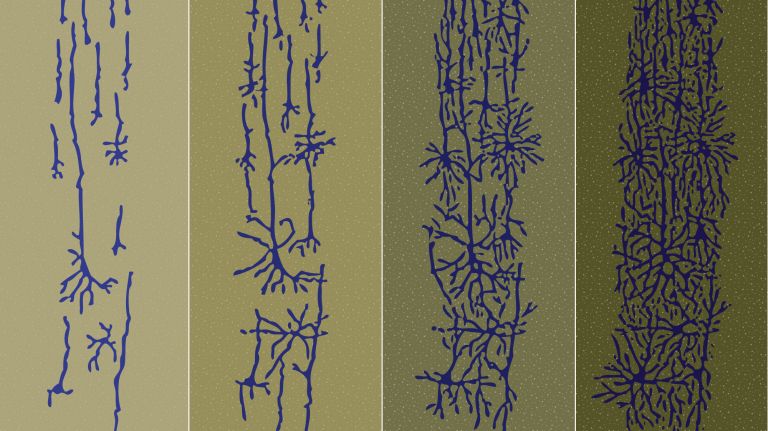
What at first glance appears to be a boring sheath turns out, upon closer inspection, to be a fascinating, dynamic, and highly complex phenomenon. Immediately after birth, much of the newborn’s brain is still a gray blur. The myelination of the axons – which gives the “white matter” in the brain and Spinal cord its name – begins during pregnancy, but continues throughout childhood and is further refined during puberty. And myelination even continues beyond that – albeit to a much lesser extent.
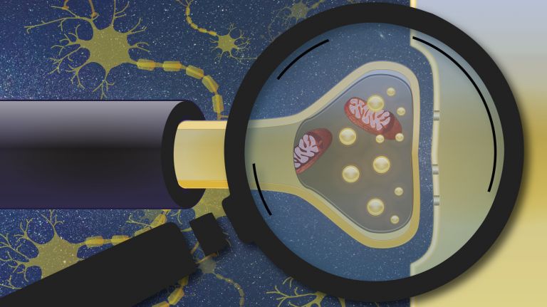
Specialized cells called Oligodendrocytes are responsible for this insulation process. Each of these cells, which belong to the glial cell family, can wrap cellular projections around several axonal segments on different axons, much like a bandage. In this way, oligodendrocytes also function as cable ties, bundling multiple axons together. They also supply the Axon and its many energy-hungry ion pumps with nutrients.
Spinal cord
medulla spinalis
The spinal cord is the part of the central nervous system located in the spine. It contains both the white matter of the nerve fibers and the gray matter of the cell nuclei. Simple reflexes such as the knee-jerk reflex are already processed here, as sensory and motor neurons are directly connected. The spinal cord is divided into the cervical, thoracic, lumbar, and sacral spinal cord.
Oligodendrocytes
Cells of the central nervous system that form the myelin sheath around nerve cells, thereby increasing their conduction velocity. They belong to the glial cells.
Axon
axon
The axon is the extension of the nerve cell that is responsible for conducting nerve impulses to the next cell. An axon can branch out many times, reaching a large number of downstream nerve cells. It can be more than a meter long. The axon ends in one or more synapses.
Speed boost through myelination
The Myelin sheaths provide a truly dramatic speed boost to the electrical signals – the action potentials – that travel along the Axon from the cell body to the synapses at the other end of the cell. Without myelin, such an Action potential propagates by gradually changing the voltage at the cell membrane along the entire length of the axon. When a certain threshold is exceeded, voltage-gated membrane channels open, allowing a sudden influx of positively charged sodium ions into the cell. As a result, the Membrane potential at this point shoots up even further, pushing the region further downstream past the threshold as well, so that the voltage-gated channels there also open. The action potential thus travels to the end of the axon via a chain reaction.
However, this process proceeds relatively slowly, at a speed of about one meter per second. Warmer temperatures or a larger axon diameter – where there is more electrically conductive internal volume per unit of membrane surface area, and thus reduced longitudinal resistance – can accelerate the journey. Some mollusks, such as squid and sea snails, use this strategy to transmit particularly fast signals via exceptionally thick axons – up to one millimeter in diameter – even without myelination.
In vertebrates, and thus also in the human brain, however, myelin ensures greater speed – while simultaneously saving a great deal of space in the highly complex central nervous system. Above all, it allows the electrical signal to simply skip over myelinated sections of the axon. Thanks to the insulating effect, the action potential generated locally can propagate relatively undisturbed as an electric field within the axon. This is sufficient to reach the threshold for opening the voltage-gated channels at the next gap where the cell membrane is exposed – the Ranvier node. The action potential now jumps at breakneck speed from one node to the next, enabling an enormous conduction speed – up to 100 meters per second.
Despite this sophisticated insulation strategy, when it comes to transmission speed, “size matters.” It depends on the thickness of the axon beneath the myelin as well as its thickness at the intervening nodes. The thickness of the myelin sheath itself and the length of the wrapped sections also play a role.
Benedikt Grothe of Ludwig Maximilian University of Munich was researching signal transmission in the auditory system, where milliseconds often make the difference in distinguishing subtle differences between sounds ▸ The Secret to Success: Communication. Through experiments and modeling, he has discovered that the fastest possible signal transmission occurs when the axon diameter is large and the myelin segments are short. However, this maximum speed comes at a cost, since with many short, isolated segments, the number of nodes along the fiber also increases – points at which the action potential must actively jump from one segment to the next – a process that requires a great deal of energy. “In practice, we therefore often observe a trade-off between speed and metabolic cost,” says Grothe. “But if the circuit demands it, the speed can be maximized.” These factors can also vary along a single axon. Toward the end of the axon, for example, the researchers observe an increasing optimization of axon diameter and myelin segment length geared toward speed. This presumably serves as a safeguard to ensure that the action potential reliably reaches the synapse.
Myelin
Myelin is a fatty substance produced by glial cells. It envelops the axons (long, fiber-like extensions) of nerve cells and insulates them, preventing messages from passing uncontrollably to neighboring nerve cells. This also greatly accelerates conduction velocity.
Axon
axon
The axon is the extension of the nerve cell that is responsible for conducting nerve impulses to the next cell. An axon can branch out many times, reaching a large number of downstream nerve cells. It can be more than a meter long. The axon ends in one or more synapses.
Action potential
In excitable cells (e.g., neurons or muscle cells), very rapid changes in electrical potential occur across the cell membrane. This event is the basis for signal conduction along the axon of the nerve cell. The action potential continues along the cell membrane and, according to the all-or-nothing principle, only occurs when the cell has been sufficiently excited.
Membrane potential
The membrane potential is a voltage measured between the inside and outside of the cell membrane. It arises from the different distribution of electrically charged particles inside and outside the cell.
Synapse
A synapse is a connection between two neurons and serves as a means of communication between them. It consists of a presynaptic region – the terminal button of the sender neuron – and a postsynaptic region – the region of the receiver neuron with its receptors. Between them lies the synaptic cleft.




Myelination as a component of plasticity
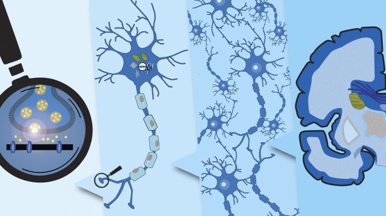
The Myelin sheath is thus more than just insulation. Rather, it is a highly precise configuration of the Axon and the surrounding Oligodendrocytes. And this configuration is by no means rigid. “Changes in myelination have much in common with neural plasticity, even if the processes take months rather than days,” says Tim Czopka, who by now moved with his research group from the Technical University of Munich to the University of Edinburgh. Together with his team, he is studying zebrafish to investigate how myelination changes in response to the activities and experiences of individual neurons. In humans, too, it can be observed that intensive and long-term learning processes affect the White matter in the brain. For example, when learning to juggle or play a new instrument, the white matter increases in certain regions; specific neural circuits become more heavily myelinated to improve signal transmission in those areas. “The connections between neurons via synapses are just one level of communication within a network, but for the impulses to arrive at the right place at the right time, myelination must also be adjusted,” says Czopka.
Once an axon is fully wrapped, it can still make some fine adjustments together with its oligodendrocytes – for example, changing its diameter or adjusting the length of the myelinated segments and the position of the myelin sheaths. However, other forms of Plasticity are then no longer possible. For example, it can no longer form axonal branches once it is encased in its “corset.” Czopka therefore suspects that the most exciting events take place wherever parts of the axon are not yet – or are no longer – myelinated. In these areas, there are many opportunities for neurons and oligodendrocytes to find their individual configuration through interaction with one another and in response to activity within the system.
Myelin
Myelin is a fatty substance produced by glial cells. It envelops the axons (long, fiber-like extensions) of nerve cells and insulates them, preventing messages from passing uncontrollably to neighboring nerve cells. This also greatly accelerates conduction velocity.
Axon
axon
The axon is the extension of the nerve cell that is responsible for conducting nerve impulses to the next cell. An axon can branch out many times, reaching a large number of downstream nerve cells. It can be more than a meter long. The axon ends in one or more synapses.
Oligodendrocytes
Cells of the central nervous system that form the myelin sheath around nerve cells, thereby increasing their conduction velocity. They belong to the glial cells.
White matter
The white matter refers to the myelinated fibers of the nervous system that connect one neuron to another. The white color is caused by the myelin sheath surrounding the fibers.
Plasticity
Neuroplasticity
The term neuroplasticity describes the ability of synapses, nerve cells, and entire areas of the brain to change structurally and functionally depending on the degree to which they are used. Synaptic plasticity refers to the adaptation of the signal transmission strength of synapses to the frequency and intensity of incoming stimuli, for example in the form of long-term potentiation or depression. In addition, the size, interconnection, and activity patterns of different areas of the brain also change depending on their use. This phenomenon is referred to as cortical plasticity when it specifically affects the cortex.
Learning from the zebrafish
Communication begins when the oligodendrocyte precursor cells go in search of partners. Inspired by signals that have largely remained unknown until now, the cells then scan their environment for suitable neurons. “They try to wrap themselves around everything they can with their stubby extensions, like little spiders,” explains Czopka, who films these processes live in the transparent larvae of zebrafish (see box). Depending on the signals they detect in the vicinity of a specific axon, they either stabilize their extensions or retract them.
It is already becoming clear that the interaction between the two cell types is very finely tuned. Oligodendrocytes and their precursors themselves have many receptors for neurotransmitters and voltage-gated ion channels, which they use to “listen” to the activity of neurons. If a nerve cell is very active, the metabolic activity of the oligodendrocytes surrounding it also changes: they divide and differentiate more rapidly. Understanding the exact nature and dynamics of these signals is one of the research goals of Czopka and his team.
Behind these research efforts lies the hope of being able to better restore the balance between neurons and oligodendrocytes in the future, after it has been disrupted – for example, by disease or injury. In ▸ multiple sclerosis, for example, misdirected immune cells attack the oligodendrocytes and destroy the Myelin sheath – at least temporarily. This jeopardizes both the neurons’ energy supply and their communication with other nerve cells. Depending on the course of the disease, however, the system can recover at least partially and regenerate destroyed myelin sheaths. The hope is that one day, through targeted intervention in the development and activity of oligodendrocytes, it will be possible to precisely repair the function of neural networks.
Oligodendrocytes
Cells of the central nervous system that form the myelin sheath around nerve cells, thereby increasing their conduction velocity. They belong to the glial cells.
Myelin
Myelin is a fatty substance produced by glial cells. It envelops the axons (long, fiber-like extensions) of nerve cells and insulates them, preventing messages from passing uncontrollably to neighboring nerve cells. This also greatly accelerates conduction velocity.
Further reading
- Ford MC et al: Tuning of Ranvier node and internode properties in myelinated axons to adjust action potential timing. Nature Communications, 2015, 6:8073 ( zum Volltext )
- Marisca R et al: Functionally Distinct Subgroups of Oligodendrocyte Precursor Cells Integrate Neural Activity and Execute Myelin Formation. Nat Neurosci. 2020 Mar; 23(3): 363–374. ( zum Volltext )