Repairing the Nervous System
In humans, injuries to the nervous system and neurodegenerative diseases usually cause permanent damage. However, we may have a latent self-healing potential that could be harnessed for future therapies.
Scientific support: Prof. Dr. Constanze Seidenbecher
Published: 30.04.2020
Difficulty: intermediate
- In humans, damage to the brain and Spinal cord does not normally regenerate. The result is permanent impairments such as paralysis or loss of cognitive abilities.
- Some animals, such as the zebrafish, have the ability to replace dead nerve cells and can thus repair damaged neural networks. Neurobiologists are trying to decipher the mechanisms behind this self-healing.
- The findings are intended to help trigger this repair process in humans as well. This could be achieved, for example, by reprogramming brain cells or introducing neural stem cells into the nervous system.
- For this to eventually work, however, we first need a deeper understanding of both the damaging processes and the regenerative capacity and functioning of neural circuits.
Spinal cord
medulla spinalis
The spinal cord is the part of the central nervous system located in the spine. It contains both the white matter of the nerve fibers and the gray matter of the cell nuclei. Simple reflexes such as the knee-jerk reflex are already processed here, as sensory and motor neurons are directly connected. The spinal cord is divided into the cervical, thoracic, lumbar, and sacral spinal cord.
Olive-brown back, pale yellow belly, zebra-like stripes on the sides. Native to the Ganges River basin, the zebrafish now also swims through numerous research laboratories. This is primarily due to a special ability: the fish can replace dead nerve cells and thus restore entire regions of the brain. In humans and other mammals, injuries to the central nervous system and neurodegenerative diseases typically lead to permanent and often severe damage, such as paralysis, speech disorders, loss of cognitive abilities, or epilepsy. It is therefore hardly surprising that researchers around the globe are interested in the fish’s nervous system and are attempting to decipher the underlying mechanisms of self-healing.
Scar-free brain
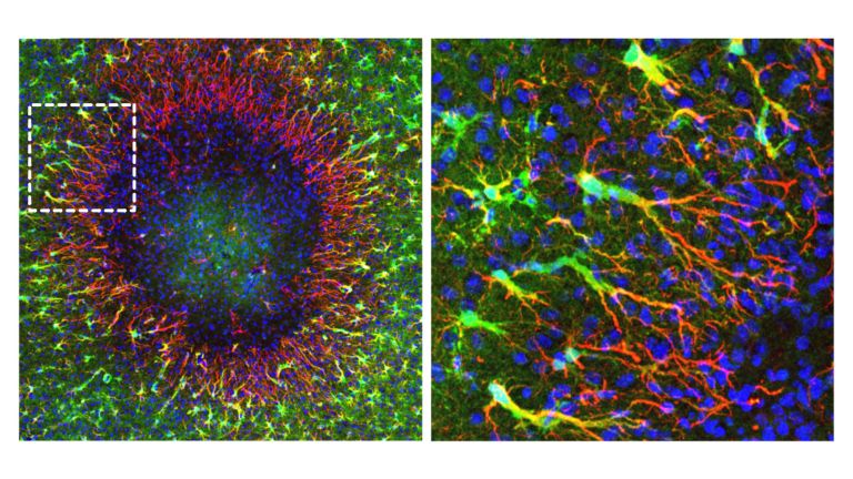
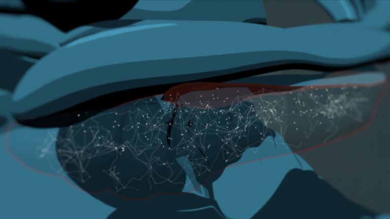
Jovica Ninkovic, group leader at the Institute for Stem Cell Research at the Helmholtz Zentrum München, is working with his team to investigate how the brains of zebrafish regenerate after puncture wounds. “It is striking that wounds close without scar tissue just a few days after the injury,” says Ninkovic. Similar to what happens in humans, so-called Glial cells gather near the wound. These are all the cells in the nervous tissue that can be structurally and functionally distinguished from nerve cells. Unlike in the human brain, however, the recruitment of glial cells in zebrafish does not lead to scar formation. The scientists’ findings suggest that this is because the types of glial cells that rush to the site for repair purposes have a very specific composition. The next step is to decipher the molecular mechanisms involved in these beneficial glial responses.
However, the absence of a scar is not enough to ensure that the damaged tissue regenerates completely. Instead, the damaged network must, so to speak, be patched back together. This is no problem for the zebrafish – nor, for that matter, for many other fish and certain species of salamanders. Throughout their lives, these animals can generate sufficient new nerve cells to provide replacement material. In this context, Ninkovic and his colleagues are investigating how the newly generated neurons contribute to the functional restoration of neural circuits.
In humans (and in mammals in general), the situation is different: It is now clear that the adult brain also possesses so-called neural stem cells. These can develop into nerve cells – a process experts refer to as adult neurogenesis. However, in adults, neural stem cells are present in only a very small number of brain regions. The purpose of adult neurogenesis has not yet been conclusively clarified. However, there is strong evidence suggesting that the integration of newly formed nerve cells into mature neural circuits occurs, if at all, only in a few specialized niches of the mammalian brain. According to current knowledge, new nerve cells are apparently rarely formed following brain injuries or in cases of neurodegenerative diseases such as Parkinson’s or Alzheimer’s. A loss of neurons usually leads to significant restructuring of existing neural networks. This sometimes reduces functional impairments, but they generally do not disappear completely.
Ninkovic hopes, however, that adult neurogenesis might be stimulated: “Among other things, we are searching for the molecular factors that are crucial for the development of certain types of nerve cells in the brains of adult mammals,” the scientist said. These findings could be used to activate corresponding molecular cascades, which in turn would induce the formation of specific types of neurons. This might make it possible to compensate for the loss of neurons following injuries or diseases.
Glial cells
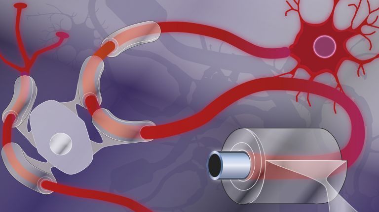
Glia cells are the second largest group of cells in the brain after neurons. For a long time, they were considered inactive elements of the brain, referred to as "nerve cement." Today, we know that the different types of glia cells (astrocytes, oligodendrocytes, and microglia in the CNS; Schwann cells in the PNS) perform clearly defined tasks in the nervous system. For example, they respond to pathogens, play an important role in nourishing nerve cells, and insulate nerve fibers. They account for slightly more than 50 percent of the brain's cells, compared to neurons.
Surprising discovery
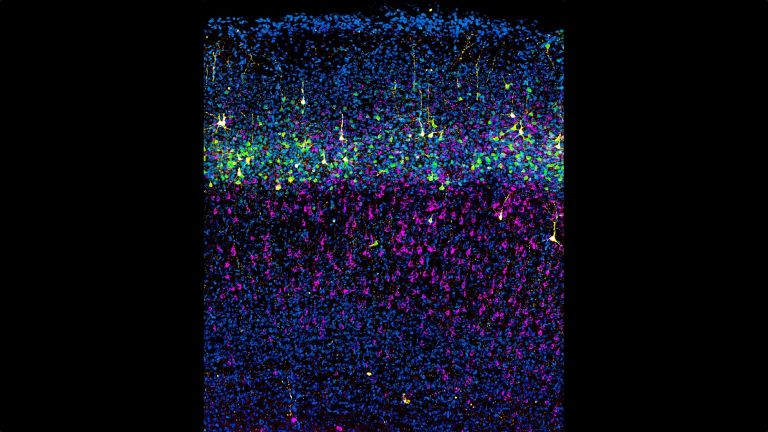
Just like her former colleague Ninkovic, neurobiologist Magdalena Götz is also researching the regeneration of the nervous system. Götz is the director of the Institute for Stem Cell Research at the Helmholtz Zentrum München and holds the Chair of Physiological Genomics at Ludwig Maximilian University of Munich. One of her most important discoveries, however, did not come from zebrafish but from experiments with mice. Around the turn of the millennium, she and her colleagues were able to show that Glial cells possess stem cell properties and can develop into nerve cells. This finding caused a minor sensation, as it contradicted the prevailing scientific consensus at the time.
“Using substances introduced into the brain, we are now trying to program glial cells to form nerve cells,” explains Götz. The team has already succeeded in doing this in mice. However, it remains to be seen whether these new nerve cells also form the correct connections and actually repair a damaged network. “We need to understand under what conditions the new nerve cells contribute meaningfully to regeneration,” says Götz. “After all, it could also be that they cause malfunctions.” That is why it is extremely important to first gain a better understanding of how neural circuits function.
The same applies to other brain repair strategies: For several years now, Götz and other researchers have been attempting to introduce new nerve cells into the brains of mice – for example, by injecting stem cells or transplanting young neurons. These new cell populations are intended to help rebuild damaged areas. Götz’s team, for instance, demonstrated that transplanted embryonic neurons can integrate into injured neural circuits within three months. The new neurons then exhibit the same functional properties as the existing ones. Such replacement therapies are conceivable in the future – at least if a sufficient portion of the existing neural network is still intact. However, a great deal of research is still needed: “Our latest experiments show that the immediate environment of the dead cells is crucial for the integration of new nerve cells,” reports Götz. “Therefore, we must first understand in detail the various types of damage in the brain – caused, for example, by a stroke or Alzheimer’s disease – in order to be able to functionally replace dead nerve cells with new ones.”
Glial cells
Glia cells are the second largest group of cells in the brain after neurons. For a long time, they were considered inactive elements of the brain, referred to as "nerve cement." Today, we know that the different types of glia cells (astrocytes, oligodendrocytes, and microglia in the CNS; Schwann cells in the PNS) perform clearly defined tasks in the nervous system. For example, they respond to pathogens, play an important role in nourishing nerve cells, and insulate nerve fibers. They account for slightly more than 50 percent of the brain's cells, compared to neurons.
stroke
Cerebral apoplexy
In a stroke, the brain or parts of it are no longer supplied with sufficient blood, which impairs the supply of oxygen and glucose. The most common cause is a blockage in an artery (ischemic stroke), less commonly a hemorrhage (hemorrhagic stroke). Typical symptoms include sudden visual disturbances, dizziness, paralysis, speech or sensory disturbances. Long-term consequences can include various sensory, motor, and cognitive impairments.
Reconnecting the spinal cord
Similar to areas of the brain, damaged Spinal cord structures normally cannot regenerate. Once the spinal cord is completely severed, it does not grow back together. Those affected remain permanently paralyzed. This is primarily due to a specific bundle of nerve fibers called the corticospinal tract. It runs from the cerebral Cortex into the spinal cord and is primarily responsible for the voluntary control of the body’s muscles.
This bundle of nerve fibers sends out more than one million neurons, which are normally established during the first two years of life. If it is severed, it generally cannot re-establish connections.
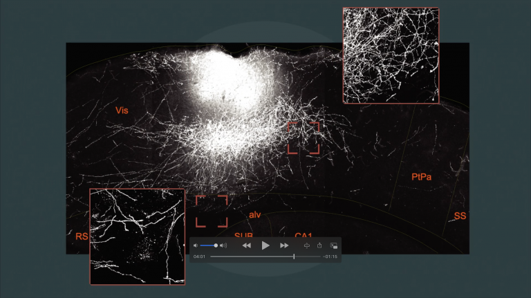
However, if residual connections remain after an injury, there is hope: “Possible regeneration depends crucially on the reorganization of existing neural circuits,” explains Florence Bareyre, head of the “Spinal Cord Repair” research group at the Institute for Clinical Neuroimmunology at Ludwig Maximilian University in Munich. For example, a severed corticospinal tract can reestablish new functional connections – in a sense, via detours – following an incomplete spinal cord injury.
Bareyre and her team are investigating how this regeneration process unfolds: Which synapses are preserved, and which are eliminated? What is necessary for new synapses to form? And how does the integration of the new networks into existing structures work? The answers to these questions could help identify ways and means to specifically stimulate this restructuring. This might make it possible to at least partially heal spinal cord injuries.
“We have already identified molecules that appear to be able to trigger the formation of new connections in the injured corticospinal tract.” The task now is to further unravel the fundamental principles of this rewiring. Bareyre is convinced that a better understanding of the underlying mechanisms would contribute significantly to the development of new therapeutic strategies for treating spinal cord injuries. So there is certainly hope that it will indeed be possible in the future to heal injuries to the nervous system – something that comes naturally to the zebrafish.
Spinal cord
medulla spinalis
The spinal cord is the part of the central nervous system located in the spine. It contains both the white matter of the nerve fibers and the gray matter of the cell nuclei. Simple reflexes such as the knee-jerk reflex are already processed here, as sensory and motor neurons are directly connected. The spinal cord is divided into the cervical, thoracic, lumbar, and sacral spinal cord.
Cortex
cortex cerebri
Cortex refers to a collection of neurons, typically in the form of a thin surface. However, it usually refers to the cerebral cortex, the outermost layer of the cerebrum. It is 2.5 mm to 5 mm thick and rich in nerve cells. The cerebral cortex is heavily folded, comparable to a handkerchief in a cup. This creates numerous convolutions (gyri), fissures (fissurae), and sulci. Unfolded, the surface area of the cortex is approximately 1,800cm².
Further reading
- Roger AB et al.: New approaches for brain repair – from rescue to reprogramming. Nature 2018 May;557(7705):329-34 (zum Abstract)
https://www.ncbi.nlm.nih.gov/pubmed/29769670 - Falkner S et al.: Transplanted embryonic neurons integrate into adult neocortical circuits. Nature 2016 Nov;539(7628):248-53 (zum Abstract).
https://www.ncbi.nlm.nih.gov/pubmed/27783592 - Grade S and Götz M: Neuronal replacement therapy: previous achievements and challenges ahead. NPJ Regen Med. 2017 Oct;23;2:29 (zum Volltext)
https://www.ncbi.nlm.nih.gov/pmc/articles/PMC5677983/ - Baumgart EV et al.: Stab wound injury of the zebrafish telencephalon: a model for comparative analysis of reactive gliosis. Glia. 2012 Mar;60(3):343-57 (zum Abstract).
https://www.ncbi.nlm.nih.gov/pubmed/22105794 - Bradley PM et al.: Corticospinal circuit remodeling after central nervous system injury is dependent on neuronal activity. J Exp Med. 2019 Aug;pii: jem.20181406. doi: 10.1084/jem.20181406. [Epub ahead of print] (zum Abstract)
https://www.ncbi.nlm.nih.gov/pubmed/31391209