
Well-Connected
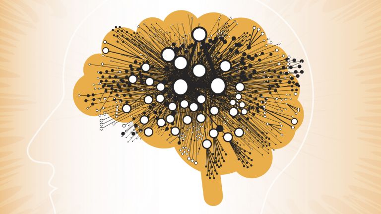
The brain consists of a complex network of nerve cells that communicate with one another. The neural circuitry is extremely efficient and constantly adapts to the challenges of life.
Scientific support: Prof. Dr. Jan Benda
Published: 30.04.2020
Difficulty: intermediate
- Parallel-processing network systems are currently the most widely accepted neuroscientific model for describing how the brain functions.
- The complex network structures in the brain are a direct result of nerve cells communicating via electrical signals.
- The networks enable the emergence of complex activity patterns through the coordinated excitation of nerve cells.
- Due to neural plasticity, the brain’s microstructure – and thus its networks – change throughout a person’s lifetime.
- After injuries, the brain regenerates using mechanisms of Plasticity and forms new networks.
Plasticity
Neuroplasticity
The term neuroplasticity describes the ability of synapses, nerve cells, and entire areas of the brain to change structurally and functionally depending on the degree to which they are used. Synaptic plasticity refers to the adaptation of the signal transmission strength of synapses to the frequency and intensity of incoming stimuli, for example in the form of long-term potentiation or depression. In addition, the size, interconnection, and activity patterns of different areas of the brain also change depending on their use. This phenomenon is referred to as cortical plasticity when it specifically affects the cortex.
“What is the body when the head is off?” William Shakespeare once asked. “Nothing!” one might be tempted to reply. After all, the brain is intrinsically linked to “being human.” Its astonishing capabilities continue to baffle philosophers, psychologists, and, above all, neuroscientists to this day. While modern analytical and imaging techniques have revealed much about the brain’s structure and how it functions, the central question remains unanswered: How are the individual physical structures organized to cope with the countless and extremely diverse demands of life? After all, the human brain, too, is ultimately based on building blocks – nerve cells – and means of communication – electrical and chemical signals – that are already present in simple organisms.
Nerve cells in synchrony
According to the most widely accepted model currently used by neuroscientists to theoretically explain how the brain functions, individual nerve cells are initially interconnected in small groups, known as modules. Several of these circuits, in turn, form larger clusters. Neuroscientists refer to the connection pathways between such structures as “pathways.” Some of the modules form particularly important nodes in the network, known as hubs.
One can thus imagine the brain as a system whose fundamental building blocks organize themselves into ensembles of various sizes, which in turn are interconnected, thereby forming networks of varying scales. Such a structure is extremely efficient, since each individual nerve cell is part of different groupings and circuits – and can therefore perform multiple tasks. If certain ensembles are not currently needed, they can be actively shut down. The networks can also operate in parallel and constantly adapt to new demands: in a sense, they are constantly evolving.
This evolution is a direct result of communication between individual nerve cells. Their “language” consists of electrical impulses, known as action potentials, which trigger excitation in the recipient. This results in activity patterns, each of which carries a specific meaning. Processes involving multiple brain regions are generally based on synchronous oscillations. These occur when entire groups of neurons fire at the same frequency – in other words, they oscillate “in sync,” so to speak. Neuroscientists assume that the brain’s complex functions are based on these recurring electrical Oscillation patterns. Both rhythmic activity within individual brain regions and activity spanning multiple regions can be observed. And the more frequently certain networks are used, the more they are strengthened.
Oscillation
Oscillations occur when many neurons fire in synchronized, rhythmic patterns. These phased fluctuations in neural activity form the basis for measurable signals in the EEG. They reflect the coordinated processing of information in the brain.
Division of labor
While the dynamic network model, including the activity patterns, explains how the brain works, it does not explain how this is physiologically implemented. It has been clear for many decades that different brain functions are anchored in different areas and structures of the brain. Experts refer to this as the principle of “functional segregation.” For example, they have discovered that the Frontal lobe significantly influences personality, social behavior, and impulsivity. The Temporal lobe is primarily responsible for hearing, speech, and memory, while the Cerebellum controls fine motor skills, among other things.
frontal
An anatomical position designation – frontal means "towards the forehead," i.e., at the front.
Frontal lobe
Lobus frontalis
The frontal cortex is the largest of the four lobes of the cerebral cortex and its functions are correspondingly comprehensive. The front area, known as the prefrontal cortex, is responsible for complex action planning (known as executive functions), which also shapes our personality. Its development (myelination) takes up to 30 years and even then is not yet complete. Other important components of the frontal cortex are Broca's area, which controls our ability to express ourselves linguistically, and the primary motor cortex, which sends movement impulses throughout the body.
Temporal lobe
Lobus temporalis
The temporal lobe is one of the four lobes of the cerebrum and is located laterally (on the side) at the bottom. It contains important areas such as the auditory cortex and parts of Wernicke's area, as well as areas for higher visual processing; deep within it lies the medial temporal lobe with structures such as the hippocampus.
Cerebellum
Cerebellum
The cerebellum is an important part of the brain, located at the back of the brain stem and below the occipital lobe. It consists of two cerebellar hemispheres covered by the cerebellar cortex and plays an important role in motor processes, among other things. It develops from the rhombencephalon.
Structured early on
As early as the third week of pregnancy, the nervous system and brain begin to form in the embryo. During this process of neurogenesis, new nerve cells develop from precursor or stem cells. These cells then build up the cerebral Cortex layer by layer; subsequently, the various brain regions develop. By the end of the eighth week of pregnancy, the basic structures of the brain and Spinal cord are almost completely in place. In the following months, vast numbers of new nerve cells form there through cell division. This basic structure of the brain remains largely unchanged throughout life.
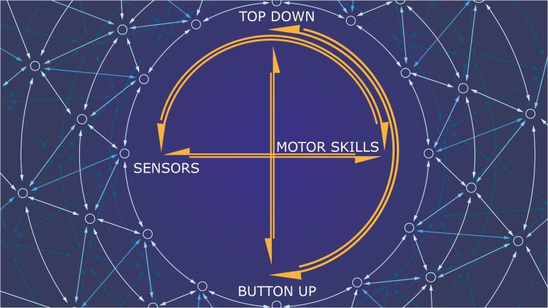
Although this basic structure is already present at birth, newborns are not yet capable of complex cognitive tasks. This is because the mere existence of the different brain regions is not sufficient to perform diverse, interdependent tasks: Actions, for example, result from thoughts and feelings or from visual and auditory impressions. The brain processes and organizes this information and then issues commands to the body. This, in turn, gives rise to new feelings, thoughts, and stimuli. These interconnections can be observed in all directions; in a sense, everything influences everything else.
Such a complex interplay can only function if no single component operates in isolation. Neuroscientists refer to this as the brain’s “functional connectivity.” There is also ample evidence for this second principle from anatomical studies of the brain: The brain’s smallest units – nerve cells, or neurons – are connected to thousands of others, for example. Experts even suspect that every single Neuron is connected to each of the approximately 86 billion other neurons in the brain via just six intermediate steps. In total, the brain is estimated to have 600 trillion neural junctions, known as synapses.
Cortex
cortex cerebri
Cortex refers to a collection of neurons, typically in the form of a thin surface. However, it usually refers to the cerebral cortex, the outermost layer of the cerebrum. It is 2.5 mm to 5 mm thick and rich in nerve cells. The cerebral cortex is heavily folded, comparable to a handkerchief in a cup. This creates numerous convolutions (gyri), fissures (fissurae), and sulci. Unfolded, the surface area of the cortex is approximately 1,800cm².
Spinal cord
medulla spinalis
The spinal cord is the part of the central nervous system located in the spine. It contains both the white matter of the nerve fibers and the gray matter of the cell nuclei. Simple reflexes such as the knee-jerk reflex are already processed here, as sensory and motor neurons are directly connected. The spinal cord is divided into the cervical, thoracic, lumbar, and sacral spinal cord.
Neuron
A neuron is a specialized cell in the nervous system that is responsible for processing and transmitting information. It receives signals via its dendrites and transmits them via its axon. Transmission occurs electrically within the neuron and, between neurons, usually chemically via synapses.
Networking
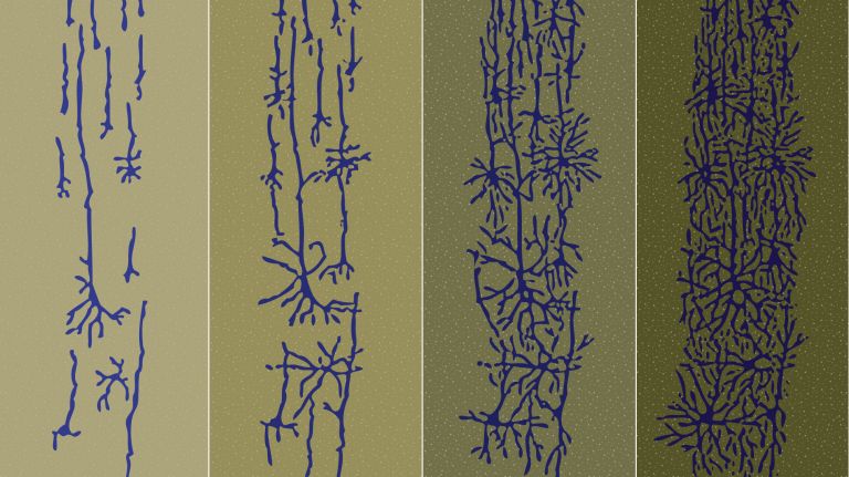
Although the majority of nerve cells are already in place in infants, this extensive network of connections is not. It is only during early childhood that the number of connections between nerve cells multiplies; in addition, the nerve fibers become significantly thicker. For these reasons, the brain’s weight triples from birth to adulthood. This process of networking is essential for absorbing, processing, and responding to the diverse information from the environment. Because of that, he first years of life, in particular, play a decisive role in the development of cognitive abilities.
The formation of these network structures originates primarily from the smallest units – the nerve cells – and is explained by Hebb’s rule: When a Neuron is repeatedly stimulated and, in turn, stimulates a neighboring neuron, this causes – to put it simply – the connection between the two to become stronger. Neurobiologists would say that the efficiency with which cell A can generate an Action potential in the neighboring cell B increases. Based on these communication processes among nerve cells, their network structures form and change. However, new connections can also be created (structural Plasticity). This ability of the brain is by no means lost after childhood but is retained throughout life.
The extent to which newly regenerating nerve cells contribute to this process has not yet been conclusively clarified. For nearly twenty years, however, it has been proven that what once seemed unthinkable is true: new nerve cells continue to form even in the adult brain – a process known as adult neurogenesis.
Neuron
A neuron is a specialized cell in the nervous system that is responsible for processing and transmitting information. It receives signals via its dendrites and transmits them via its axon. Transmission occurs electrically within the neuron and, between neurons, usually chemically via synapses.
Action potential
In excitable cells (e.g., neurons or muscle cells), very rapid changes in electrical potential occur across the cell membrane. This event is the basis for signal conduction along the axon of the nerve cell. The action potential continues along the cell membrane and, according to the all-or-nothing principle, only occurs when the cell has been sufficiently excited.
Plasticity
Neuroplasticity
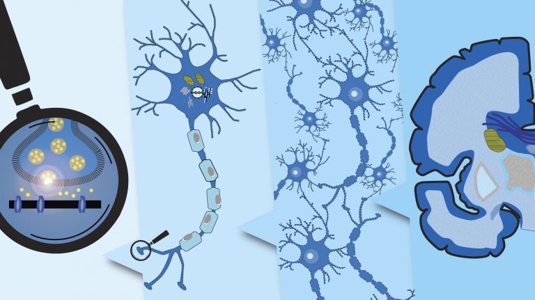
The term neuroplasticity describes the ability of synapses, nerve cells, and entire areas of the brain to change structurally and functionally depending on the degree to which they are used. Synaptic plasticity refers to the adaptation of the signal transmission strength of synapses to the frequency and intensity of incoming stimuli, for example in the form of long-term potentiation or depression. In addition, the size, interconnection, and activity patterns of different areas of the brain also change depending on their use. This phenomenon is referred to as cortical plasticity when it specifically affects the cortex.



Getting in shape
Without neural plasticity, we humans would be unable to master life’s ever-changing demands: As soon as we learn something new – whether it is a word, a mathematical operation, or a dance step – the connections between nerve cells, in particular, change. And to a certain extent, larger structures do as well. This is particularly evident in people who regularly engage in an activity and have therefore mastered it exceptionally well. In professional musicians, certain brain regions are more highly developed compared to non-musicians. And depending on their training – for example, as string players, drummers, or conductors – distinct characteristics can be identified in their brains. Brain structure is thus shaped by years of training. In addition, studies show that entire brain regions can also completely reorient themselves: In people who have been deaf since birth, for example, the previously unused part of the cerebral Cortex is assigned new tasks. A similar phenomenon can be observed in people who have undergone limb amputations.
Cortex
cortex cerebri
Cortex refers to a collection of neurons, typically in the form of a thin surface. However, it usually refers to the cerebral cortex, the outermost layer of the cerebrum. It is 2.5 mm to 5 mm thick and rich in nerve cells. The cerebral cortex is heavily folded, comparable to a handkerchief in a cup. This creates numerous convolutions (gyri), fissures (fissurae), and sulci. Unfolded, the surface area of the cortex is approximately 1,800cm².
Repair in the Brain
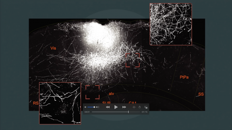
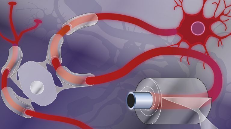
Despite the impressive adaptability of nerve cells, the brain has a harder time repairing injuries compared to other tissues. To a certain extent, it achieves this by forming new connections between nerve cells that are still functional ▸ Repairing the Nervous System. A massive and well-coordinated reorganization then takes place in the affected regions.
However, it is unclear to what extent damaged or destroyed brain cells can be replaced by new ones. Regeneration in the brain likely occurs primarily through existing nerve cells taking over the tasks of the lost neurons. This probably follows the fundamental principles that also apply to the formation of neural circuits during brain development and in learning processes. In a sense, then, it is particularly the adaptable and malleable network structures that define the uniqueness of the human brain. However, it is rather unlikely that Shakespeare was already alluding to this neural networking in the “head.”
Further reading
- >Van den Heuvel MP, Sporns O, Network hubs in the human brain, Trends Cogn Sci, 2013 Dec;17(12):683-696 (full text).
- Sporns O, Chialvo DR, et al., Organization, development, and function of complex brain networks, Trends Cogn Sci, 2004 Sep;8(9):418-425 ( to the abstract )
- Sporns O, Contributions and challenges for network models in cognitive neuroscience; Nat Neurosci. 2014 May;17(5):652-660 ( to the abstract )