The Fate of a Cell
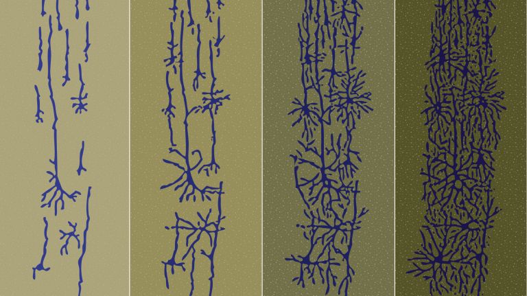
A single fertilized egg cell gives rise to one of the most complex structures in existence – the human brain. This requires a great deal of folding and numerous critical steps.
Scientific support: Dr. Ruth Beckervordersandforth-Bonk
Published: 30.04.2020
Difficulty: intermediate
- In the embryo, everything becomes increasingly diverse. Before the nervous system begins to develop, its precursors have already made numerous critical decisions and embarked on different paths through complex folding and signaling processes.
- Nerve cells arise from the ectoderm, the outermost cell layer of the embryo. During neurulation, it folds into the neural tube, the precursor of the brain and spinal cord.
- Most neurons have a very specific type of stem cell as their precursor: radial Glial cells These cells can give rise not only to nerve cells and other neuronal precursors, but also to other glial cells. Young neurons also use their long processes to make their way into the outer layers of the brain.
- The type of nerve cell into which a new Neuron will develop is determined soon after its final division. Already within the neural tube, signaling molecules are distributed in varying concentrations along the axes, thereby determining the fate of the cells.
- Learning and migration days: Most neurons migrate over long distances from their birthplace deep within the brain to their destinations. Once they arrive, they send their growing Axon on its journey.
- Young neurons are aided on their way by the growth Cones at the tips of their extensions. These serve as both sensory and locomotor organs and later transform into the synaptic terminals of the dendrites and the axon.
Glial cells
Glia cells are the second largest group of cells in the brain after neurons. For a long time, they were considered inactive elements of the brain, referred to as "nerve cement." Today, we know that the different types of glia cells (astrocytes, oligodendrocytes, and microglia in the CNS; Schwann cells in the PNS) perform clearly defined tasks in the nervous system. For example, they respond to pathogens, play an important role in nourishing nerve cells, and insulate nerve fibers. They account for slightly more than 50 percent of the brain's cells, compared to neurons.
Neuron
A neuron is a specialized cell in the nervous system that is responsible for processing and transmitting information. It receives signals via its dendrites and transmits them via its axon. Transmission occurs electrically within the neuron and, between neurons, usually chemically via synapses.
Axon
axon
The axon is the extension of the nerve cell that is responsible for conducting nerve impulses to the next cell. An axon can branch out many times, reaching a large number of downstream nerve cells. It can be more than a meter long. The axon ends in one or more synapses.
Cones
The cones are a type of photoreceptor in the retina. The three different types of cones – S, M, and L – are each stimulated by short, medium, and long wavelengths of visible light, enabling color vision. They are highly concentrated in the fovea and enable sharp vision.
It all begins with the fertilized egg. Almost everyone has seen photos in which it rests in the frame, round and uniform, like a giant moon. It’s hard to imagine that this unassuming structure contains all the information for a fateful dance so complex that, even with the combined efforts of biology to date, scientists have not yet succeeded in fully deciphering its choreography. The crowning achievement of this masterpiece: the formation of the brain. The intricate way it folds into itself and weaves networks of trillions of synapses would make origami experts and master lace-makers alike green with envy.
But back to the beginning: The uniformity of the zygote is already lost during the course of the first cell divisions. Differences in the distribution of signaling molecules – which are initially barely measurable and arise either randomly or due to the internal architecture of the cell – gradually intensify, aided by external stimuli, such as when cells come into contact with one another. Soon, the embryo’s cells set off on different paths. Only a small fraction of them will become the baby; the rest form parts of the placenta and the amniotic membranes.
The most important time in life
The cells of the embryo itself initially organize themselves into two layers – until some cells from the outer layer migrate toward the center. This first major folding process, called gastrulation, gives rise to the three germ layers: the ectoderm, mesoderm, and endoderm. Together, they form the building blocks for all subsequent organ development. “Neither birth, marriage, nor death, but gastrulation is the truly most important time in your life,” developmental biologist Lewis Wolpert once said.
The nervous system develops from cells of the ectoderm. And this process, known as neurulation, also begins with the art of folding: In the initially uniform layer of columnar cells, a groove forms in the center, which soon deepens into a trench with raised edges. The edges move toward each other until they meet in the center and fuse – the neural tube has formed, the precursor to the brain and Spinal cord In its anterior region, three outpouchings – the cerebral vesicles – soon form. They will later become the forebrain, midbrain, and hindbrain. Even in a cross-section of the neural tube, signs of change are evident. In response to signaling molecules that are distributed differently from top to bottom, various genetic programs are activated in the cells along the dorsal-ventral axis. At the very bottom, for example, the floor plate – together with even deeper-lying mesodermal cells – produces the molecule sonic hedgehog, named after a video game character. Its concentration, in a complex interplay with other molecules, determines the fate of the cells in the neural tube.
Spinal cord
medulla spinalis
The spinal cord is the part of the central nervous system located in the spine. It contains both the white matter of the nerve fibers and the gray matter of the cell nuclei. Simple reflexes such as the knee-jerk reflex are already processed here, as sensory and motor neurons are directly connected. The spinal cord is divided into the cervical, thoracic, lumbar, and sacral spinal cord.
Video game character with a formative role
The farther the cells are from the basal plate, the less sonic hedgehog reaches them, and they develop differently as a result. The cells closest to the basal plate, roughly at the level of the developing spinal cord, become interneurons, which serve as switching elements between two or more nerve cells. Next come motor neurons, which extend to the muscles and control their movements; followed by three additional layers of different Interneurons. In the upper part of the neural tube, other signaling molecules secreted by cells in the roof plate also orchestrate a fine-tuned differentiation. This leads to the formation of various sensory neurons. The basic structure of the future Spinal cord is thus already recognizable long before the first neurons differentiate. This pattern formation also takes place in the developing brain. However, it is more complex and variable due to the bends, protrusions, and indentations in the various brain regions.
Regardless of such regional differences, the birth and maturation of young nerve cells everywhere follow a similar three-act process: First comes cell proliferation. When the neural tube begins to form in the third week of development, the embryo is only a few millimeters in size. That’s not enough for much of a brain. In the weeks that follow, the production of neural progenitor cells therefore proceeds at a rapid pace. According to current estimates, approximately 4.6 million cells are produced per hour in the neuroectoderm at the peak of neurogenesis. Starting in the fifth week of development, the neuroepithelial cells in the neural tube gradually transform into radial glial cells.
Interneurons
A small multipolar neuron that is neither sensory nor motor, connected between two other neurons and conducting impulses from one nerve cell to another. In the central nervous system, interneurons are mostly inhibitory and use the neurotransmitters GABA and glycine.
Spinal cord
medulla spinalis
The spinal cord is the part of the central nervous system located in the spine. It contains both the white matter of the nerve fibers and the gray matter of the cell nuclei. Simple reflexes such as the knee-jerk reflex are already processed here, as sensory and motor neurons are directly connected. The spinal cord is divided into the cervical, thoracic, lumbar, and sacral spinal cord.
From helper to source of new neurons
Until the turn of the millennium, radial Glial cells were considered merely “helpers” whose cable-like processes guide young neurons to their destinations. But we now know that radial glial cells function as stem cells in the developing brain. This means that most neurons descend directly or indirectly from them. For a long time, hardly anyone was willing to believe this. “The most difficult part of such an unconventional finding is not the data collection, but breaking with an established dogma,” writes Munich-based neurobiologist Magdalena Götz, who was involved in the discovery of the neurogenic function of radial glial cells.
Radial glial cells divide on the inner wall of the neural tube. One of the daughter cells remains a radial glial cell. The other develops either directly into a Neuron or into another type of progenitor cell, from which neurons develop later. However, radial glial cells can also divide symmetrically into two neurons or progenitor cells – or they can mature into other types of glial cells. Which path precursor cells take depends on the region of the nervous system and the stage of development, as well as on the specific signaling cocktails associated with each. Maturation into glial cells, for example, occurs primarily later in development, when neurogenesis is largely complete. And some neurons, such as those in the spinal cord, arise directly from neuroepithelial cells without passing through the intermediate stage of the radial glial cell.
Glial cells
Glia cells are the second largest group of cells in the brain after neurons. For a long time, they were considered inactive elements of the brain, referred to as "nerve cement." Today, we know that the different types of glia cells (astrocytes, oligodendrocytes, and microglia in the CNS; Schwann cells in the PNS) perform clearly defined tasks in the nervous system. For example, they respond to pathogens, play an important role in nourishing nerve cells, and insulate nerve fibers. They account for slightly more than 50 percent of the brain's cells, compared to neurons.
Neuron
A neuron is a specialized cell in the nervous system that is responsible for processing and transmitting information. It receives signals via its dendrites and transmits them via its axon. Transmission occurs electrically within the neuron and, between neurons, usually chemically via synapses.
Young neurons on the move
Once neurons are born, they no longer divide. However, they often still have a long way to go before they reach their destination and assume their mature form and function. And so begins the second phase of maturation. From their birthplace deep in the center of the brain, near the ventricles – where the inner wall of the neural tube once lay – the new neurons migrate out into the vast world of the growing brain. In doing so, they often trail along the long processes of their neighboring radial Glial cells The first neurons have the shortest path, since their cell bodies form the inner layers of the brain. The “youngest siblings,” on the other hand, must travel farther. They pass by their earlier-born siblings into regions of the brain that lie farther and farther out. Many cells also have to cover additional distances to reach their destination – forward, backward, left, or right. Signal molecules – secreted by already established nerve and glial cells or by other tissues – serve as signposts for these young pioneers.
Visually speaking, fresh neurons initially have little in common with their mature counterparts. They resemble Teletubbies or Minions: Instead of an axon, they have several shorter extensions, called neurites, one of which eventually develops into an axon, while others mature into dendrites or are reabsorbed. Internally, however, the young nerve cells are already much more specialized than their immature exterior would suggest. “The fate of the Neuron – exactly what type of cell it will become – is determined soon after it exits the cell cycle,” says Leanne Godinho of the Technical University of Munich, who studies the maturation of nerve cells in zebrafish. “So the cells by no means set off naively and only determine their identity along the way or after arrival.” Instead, the cells receive a large portion of the identity-defining signals already in the neural tube, when the distribution of signaling molecules such as sonic hedgehog sketches out basic fate patterns with broad brushstrokes. Although these initial settings do not yet determine the exact fate of the cells, they make young neurons receptive to different signaling molecules – and thus guide them onto their respective specific migration paths.
Glial cells
Glia cells are the second largest group of cells in the brain after neurons. For a long time, they were considered inactive elements of the brain, referred to as "nerve cement." Today, we know that the different types of glia cells (astrocytes, oligodendrocytes, and microglia in the CNS; Schwann cells in the PNS) perform clearly defined tasks in the nervous system. For example, they respond to pathogens, play an important role in nourishing nerve cells, and insulate nerve fibers. They account for slightly more than 50 percent of the brain's cells, compared to neurons.
Neuron
A neuron is a specialized cell in the nervous system that is responsible for processing and transmitting information. It receives signals via its dendrites and transmits them via its axon. Transmission occurs electrically within the neuron and, between neurons, usually chemically via synapses.
Act three of the epic: partner search and networking
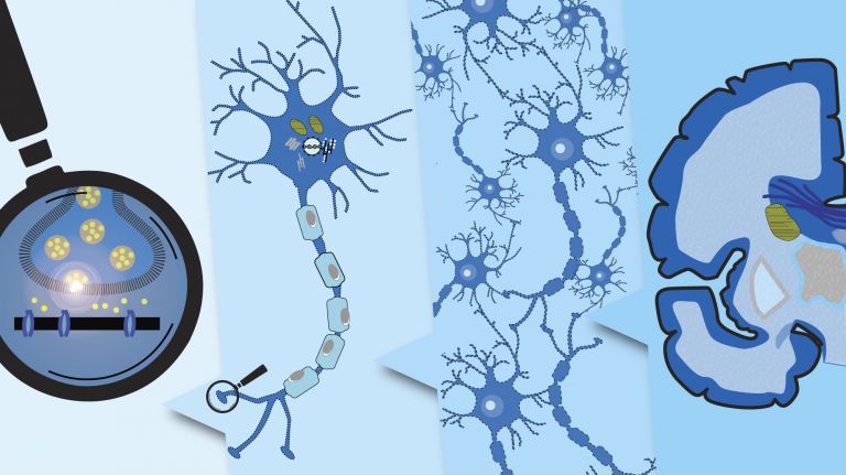
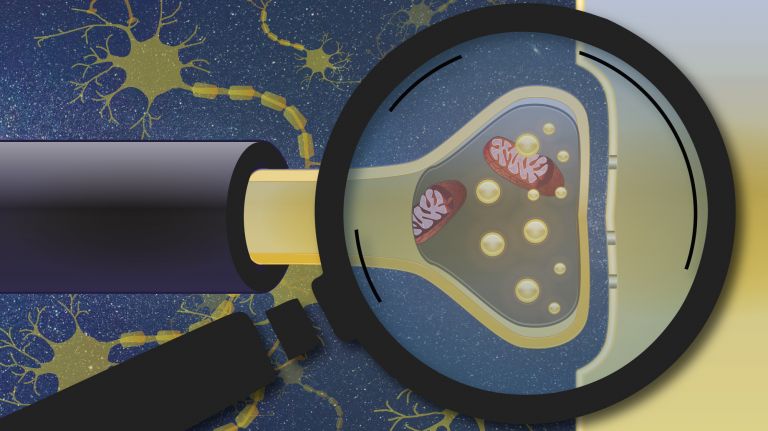
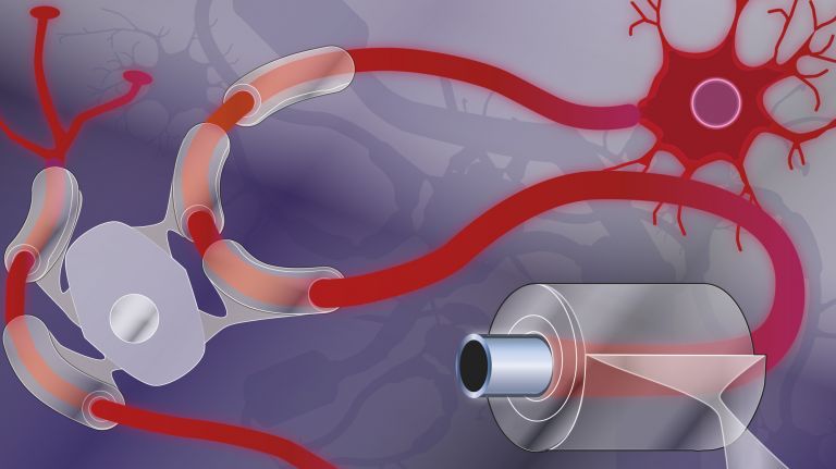
Once the cell body has reached its destination, the third act begins, in which the cell grows into its final form and establishes connections: the Axon and dendrites form and set out to find partners. Initially, all neurites of a new nerve cell have what is known as a growth cone at their tip. It functions as both a sensory and locomotor organ. Its membrane contains numerous receptors that detect and integrate physical, chemical, and electrical signals from the environment. Within the cell plasma of the cone, an abundance of skeletal and motor molecules, as well as a plentiful supply of energy-producing mitochondria, ensure that the neurite can respond to these signals – for example, by moving forward or veering off course. In particular, the growth cone of the budding axon leads an immense growth column in its wake: human axons can grow up to one meter in length.
At the end of their exploratory movements, the tips of the axon and dendrites connect to other nerve cells via synapses. In the axon, the growth Cones transform into presynaptic terminals, through which a Neuron transmits signals into the synaptic cleft. In dendrites, the growth cone becomes dendritic spines, through which the cell receives signals from synapses. The cell’s fate is now – at least in broad terms – sealed, and the great dance of development is over.
Axon
axon
The axon is the extension of the nerve cell that is responsible for conducting nerve impulses to the next cell. An axon can branch out many times, reaching a large number of downstream nerve cells. It can be more than a meter long. The axon ends in one or more synapses.
Cones
The cones are a type of photoreceptor in the retina. The three different types of cones – S, M, and L – are each stimulated by short, medium, and long wavelengths of visible light, enabling color vision. They are highly concentrated in the fovea and enable sharp vision.
Neuron
A neuron is a specialized cell in the nervous system that is responsible for processing and transmitting information. It receives signals via its dendrites and transmits them via its axon. Transmission occurs electrically within the neuron and, between neurons, usually chemically via synapses.
No end in sight – but things are calming down
The drama, however, continues throughout life ▸ Well-connected. The brain continues to develop long after birth. Synapses are first formed in large numbers and later – especially during puberty – selectively pruned to fine-tune neural networks ▸ The Nervous System – a Master of Adaptation. Myelin cells coat neurons to make signal transmission more efficient. And even new nerve cells are occasionally formed, migrate, and mature in the adult brain: in the nose, where olfactory cells regrow throughout life, and in the hippocampus, where new memories are processed. “This is no longer about rapidly producing huge numbers of neurons to build an entire brain, but rather about preserving developmental potential,” says Jovica Ninkovic of the Helmholtz Zentrum München, who is investigating regenerative capabilities in the adult brain. In his experiments, he succeeded in demonstrating that neuronal stem cells in the adult mouse brain are “frozen” in their development by different molecular mechanisms than those in the fetus. These mechanisms could react particularly sensitively to inflammation or injury in the brain – thereby providing fresh neurons in a targeted manner when the need arises.
Whether, how, and to what extent adult neurogenesis also occurs in humans is still a matter of debate, but there is enormous interest in regenerative medicine ▸ Repairing the Nervous System. “The odds are good that we can learn to activate neurogenesis even in places where it might not occur on its own,” says Ninkovic. The team led by Magdalena Götz demonstrated, for example, that embryonic neurons can successfully carry out their developmental program even after being transplanted into an adult brain. Leanne Godinho, too, hopes for a major breakthrough: “If we succeed in applying the findings from developmental biology to regenerative medicine, we might one day be able to replace neurons destroyed by injury or disease and restore function.”
Adaptation
Adaptation refers to the process by which the sensory organs, the perceptual system, or the entire organism adjusts to the intensity and quality of stimuli and to changes in environmental conditions. In visual adaptation, for example, the pupil and the sensitivity of the photoreceptors regulate themselves according to the prevailing light conditions.
Myelin
Myelin is a fatty substance produced by glial cells. It envelops the axons (long, fiber-like extensions) of nerve cells and insulates them, preventing messages from passing uncontrollably to neighboring nerve cells. This also greatly accelerates conduction velocity.
Further reading
- Silbereis, John C et al.: The Cellular and Molecular Landscapes of the Developing Human Central Nervous System. Neuron 2016; 89(2): 248–268. (Full text)
- Paridaen, Judith TML and Huttner, Wieland B: Neurogenesis during development of the vertebrate central nervous system. EMBO Report 2014; 15(4): 351–364. (Full text)
- Online embryology course for medical students. Developed by the Universities of Fribourg, Lausanne, and Bern (Switzerland). (Website)
Neuron
A neuron is a specialized cell in the nervous system that is responsible for processing and transmitting information. It receives signals via its dendrites and transmits them via its axon. Transmission occurs electrically within the neuron and, between neurons, usually chemically via synapses.