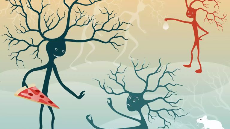
The Fear Circuit
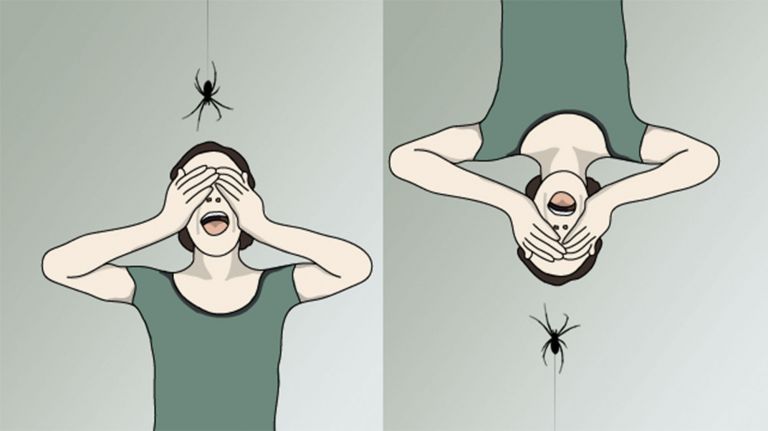
The sight of a spider or a shadow darting across the dark triggers the brain's sensitive alarm system in a flash, resulting in sweating and sheer terror. Often, it is a false alarm. But the brain quickly corrects itself.
Scientific support: Prof. Dr. Alfons Hamm
Published: 11.09.2025
Difficulty: intermediate
- The Amygdala assesses dangers and controls the cascade of fear responses.
- It receives a rough outline of the situation directly from the thalamus in order to quickly assess the danger.
- A more detailed analysis is provided a little later by the slower route from the thalamus via the Neocortex and the hippocampus.
Amygdala
corpus amygdaloideum
An important core area in the temporal lobe that is associated with emotions: it evaluates the emotional content of a situation and reacts particularly to threats. In this context, it is also activated by pain stimuli and plays an important role in the emotional evaluation of sensory stimuli. Inaddition, it is involved in linking emotions with memories, emotional learning ability, and social behavior. The amygdala is part of the limbic system.
Neocortex
The neocortex is the phylogenetically youngest part of the cerebral cortex. Since it is structured relatively uniformly in six layers, it is also referred to as the isocortex.
Hippocampus
The hippocampus is the largest part of the archicortex and an area in the temporal lobe. It is also an important part of the limbic system. Functionally, it is involved in memory processes, but also in spatial orientation and learning. It comprises the subiculum, the dentate gyrus, and the Ammon's horn with its four fields CA1-CA4.
Changes in the structure of the hippocampus due to stress are associated with chronic pain. The hippocampus also plays an important role in the amplification of pain through anxiety.
“Is emotion a magical product,” asked neuroanatomist James Papez (1883–1958) in 1937, “or is it a physiological process which depends on an anatomical mechanism?” Papez opted for the second answer. He believed that emotion was a product of the limbic system, which physician Paul Broca (1824–1880) had already described in 1878 as the “grand lobe limbique”. This phylogenetically ancient area consists of several connected structures, including the amygdala, the hippocampus, and the Septum. However, anatomists still argue today about which structures exactly belong to the Limbic system and whether it can even be called a “system”. This is because some of the components of the limbic lobe play a role in Emotions and sexual behavior, while others are responsible for memory, motivation, and navigation.
Septum
area septalis
The septal nuclei are located medially in the basal forebrain, near the anterior tip of the cingulate gyrus. They are connected to the olfactory cortex and linked to other limbic structures via the fornix. Functionally, they play a role in emotional processes and reward processing.
Limbic system
The limbic system is a functional unit in the brain. It consists of interconnected structures, primarily in the cerebrum and diencephalon. The structures assigned to the system vary depending on the source, but the most important components are the hippocampus, amygdala, cingulate gyrus, septum, and mammillary bodies. The limbic system is involved in autonomic and visceral processes as well as in mechanisms of emotion, memory, and learning. Some authors mistakenly reduce the limbic system to the emotional world by referring to it as the "emotional brain."
Emotions
Neuroscientists understand "emotions" to be complex response patterns that include experiential, physiological, and behavioral components. They arise in response to personally relevant or significant events and generate a willingness to act, through which the individual attempts to deal with the situation. Emotions typically occur with subjective experience (feeling), but differ from pure feeling in that they involve conscious or implicit engagement with the environment. Emotions arise in the limbic system, among other places, which is a phylogenetically ancient part of the brain. Psychologist Paul Ekman has defined six cross-cultural basic emotions that are reflected in characteristic facial expressions: joy, anger, fear, surprise, sadness, and disgust.
The assumption that emotion and rationality are spatially separated in the brain is widespread among laypeople. Many believe that Emotions are located in the right Hemisphere and reason in the left. In fact, the right hemisphere appears to be particularly important for processing emotions. After right-sided brain injuries, patients find it more difficult to interpret emotions in other people's faces. However, injuries to the left hemisphere also affect the emotional world: patients often suffer from a so-called catastrophic reaction with deep Depression. This suggests that the left hemisphere brightens our emotional state by inhibiting the right hemisphere. Studies with newborns also suggest that the left hemisphere is more active in positive emotions, while the right hemisphere is more active in negative emotions.
However, neuroscientists warn against attributing complex phenomena such as emotions to a single hemisphere of the brain. This is because both hemispheres are involved in almost all functions, albeit to varying degrees. In split-brain patients, whose neural connection between the two hemispheres has been severed, the relative strengths of the two brain hemispheres can be clearly observed: the left hemisphere is better at searching for causes and explanations. In contrast, the right hemisphere has a special talent for spatial-visual tasks.
Emotions
Neuroscientists understand "emotions" to be complex response patterns that include experiential, physiological, and behavioral components. They arise in response to personally relevant or significant events and generate a willingness to act, through which the individual attempts to deal with the situation. Emotions typically occur with subjective experience (feeling), but differ from pure feeling in that they involve conscious or implicit engagement with the environment. Emotions arise in the limbic system, among other places, which is a phylogenetically ancient part of the brain. Psychologist Paul Ekman has defined six cross-cultural basic emotions that are reflected in characteristic facial expressions: joy, anger, fear, surprise, sadness, and disgust.
Hemisphere
The cerebrum and cerebellum each consist of two halves – the right and left hemispheres. In the cerebrum, they are connected by three pathways (commissures). The largest commissure is the corpus callosum.
Depression
A mental illness whose main symptoms are sadness and a loss of joy, motivation, and interest. Current classification systems distinguish between different types of depression.
The hormonal and vegetative reactions to the feeling of fear and the automatic behavioral programs that follow serve to ensure survival. By releasing stress hormones, the pituitary gland enables the threatened person to act more quickly and efficiently. The basal forebrain additionally increases alertness and arousal. The autonomic system is activated via the Brain stem blood pressure and the frequency of breathing and heartbeat increase, muscles contract – the frightened person is ready to flee or fight. To prevent injuries from distracting humans or animals, the brain stem also reduces pain Perception. Finally, the brain stem also provides automated behavioral programs, such as Facial expressions of fear or cries of fear, to call for help from others.
Brain stem
truncus cerebri
The "trunk" of the brain, to which all other brain structures are "attached," so to speak. From bottom to top, it comprises the medulla oblongata, the pons, and the mesencephalon. It transitions into the spinal cord below. It is a center for vital functions such as breathing and heartbeat and contains ascending and descending pathways between the cerebrum, cerebellum, and spinal cord.
Perception
The term describes the complex process of gathering and processing information from stimuli in the environment and from the internal states of a living being. The brain combines the information, which is perceived partly consciously and partly unconsciously, into a subjectively meaningful overall impression. If the data it receives from the sensory organs is insufficient for this, it supplements it with empirical values. This can lead to misinterpretations and explains why we succumb to optical illusions or fall for magic tricks.
Facial expressions
Five muscle groups control the visible movements on the surface of our faces – and this applies to everyone in the world. Neuroscientists emphasize universal, evolutionarily anchored reactions as the reason for this. For this reason, the basic emotions of fear, anger, disgust, sadness, surprise, and joy leave similar traces on the face everywhere, which we can usually identify reliably even in strangers.
“It was just before midnight,” reports 48-year-old Martha Kristensen, "my husband and I were standing in front of our hotel in Naples. Suddenly, someone kicked me in the back of the knee from behind, threw me to the ground, and grabbed me and my backpack. I was terrified that they would kidnap me and drag me into a car. My heart was racing, I was frozen with fear. It happened so fast that I couldn't even scream for help."
Source of emotions
Fear: a feeling that everyone knows, even if they have been spared terrible experiences like that of architect Martha Kristensen (name changed by the editors). We are afraid of heights, plane crashes, dogs, our boss. When we feel this emotion, we feel it from the tips of our toes to the top of our heads. Our heart beats faster, our hands tremble, we sweat, our stomach rumbles.
Scientists have long been interested in the structures in the brain that cause us to freeze in fear or anxiety. Numerous studies on animals and humans have been conducted over the past decades to answer this question. And so it comes as no surprise that the mechanisms of fear are now among the most thoroughly researched circuits of our emotional apparatus.
It is now proven that one structure in particular plays a major role in this process: the Amygdala (almond kernel). It is part of the limbic system, which is believed to play an important role in emotion processing (see info box). The amygdala also plays a central role in aggression. It consists of two almond-shaped clusters of nerve cell bodies located in the center of the human brain, one in the left and one in the right temporal lobe, directly in front of the hippocampus.
Even the smallest injuries to the structures of the amygdala are enough to completely change an animal's behavior: “Wild-caught birds,” reported biologist Richard Phillips of the Virginia Polytechnic Institute, “which normally try to escape in panic, suddenly become completely calm.” Laboratory rats with a Lesion of the amygdala curiously explore sedated cats. Conversely, electrical stimulation of small cell clusters in the amygdala is enough to make cats cower fearfully from mice or react angrily with raised fur, hunched backs, and dilated pupils – depending on which area of the amygdala was stimulated.
Amygdala
corpus amygdaloideum
An important core area in the temporal lobe that is associated with emotions: it evaluates the emotional content of a situation and reacts particularly to threats. In this context, it is also activated by pain stimuli and plays an important role in the emotional evaluation of sensory stimuli. Inaddition, it is involved in linking emotions with memories, emotional learning ability, and social behavior. The amygdala is part of the limbic system.
Hippocampus
The hippocampus is the largest part of the archicortex and an area in the temporal lobe. It is also an important part of the limbic system. Functionally, it is involved in memory processes, but also in spatial orientation and learning. It comprises the subiculum, the dentate gyrus, and the Ammon's horn with its four fields CA1-CA4.
Changes in the structure of the hippocampus due to stress are associated with chronic pain. The hippocampus also plays an important role in the amplification of pain through anxiety.
Lesion
A lesion is damage to organic tissue.
Innate and learned fears
The Amygdala thus serves as an alarm system for animals and humans. Within a few milliseconds, it evaluates situations and assesses dangers. Some sights, sounds, or smells trigger fear from birth or after a single encounter. For example, laboratory rats that have never lived in the wild are afraid when they hear the cry of an owl or smell a predator.
Some fears are not innate, but very easy to acquire. Monkeys, for example, fear snakes as soon as they observe another monkey reacting with fear to a reptile. The amygdala of primates reacts similarly sensitively to negative Facial expressions of others. Evolutionarily, such innate fears or tendencies toward fear are of great advantage to the individual creature: for example, if a rat quickly flees when it hears an owl hooting, it may save its life.
However, even stimuli that have long been perceived as neutral or positive can become associated with danger through learning processes and later trigger fear themselves. If a neutral stimulus occurs simultaneously or shortly before an unpleasant stimulus such as pain, the fear triggered by the unpleasant stimulus rubs off on the neutral stimulus. The sounds Martha Kristensen heard immediately before she was kicked in the back of the knee were stored by her amygdala as threatening. “When I hear footsteps behind me today,” she says, “especially at night, I still feel afraid. I turn around or walk faster.”
Amygdala
corpus amygdaloideum
An important core area in the temporal lobe that is associated with emotions: it evaluates the emotional content of a situation and reacts particularly to threats. In this context, it is also activated by pain stimuli and plays an important role in the emotional evaluation of sensory stimuli. Inaddition, it is involved in linking emotions with memories, emotional learning ability, and social behavior. The amygdala is part of the limbic system.
Facial expressions
Five muscle groups control the visible movements on the surface of our faces – and this applies to everyone in the world. Neuroscientists emphasize universal, evolutionarily anchored reactions as the reason for this. For this reason, the basic emotions of fear, anger, disgust, sadness, surprise, and joy leave similar traces on the face everywhere, which we can usually identify reliably even in strangers.


Circuitry of fear
But how does the brain actually know whether a situation is dangerous? Neuroscientist Joseph LeDoux of New York University has described the underlying mechanisms as a fear circuit that sends information to the Amygdala in two ways: one fast, rough, and error-prone, and one slow but verified by precise analysis.
The starting point is always the thalamus. This part of the Diencephalon forms the gateway to consciousness and is an important central hub for messages from the sensory organs. When it receives an emotional stimulus, such as a loud noise, it forwards a rough sketch of the sensory impression directly to a small group of cells (“fear-on” neurons) in the lateral amygdala.
When these cell clusters are activated, the information flows on to the central core of the amygdala. This is where the defensive behavior programs are activated. This triggers physical fear responses, as Martha Kristensen describes: “Everything happened so fast, I was afraid, my heart was racing, I was frozen with fear. I only felt the many bruises afterwards.” Thanks to this thalamo-amygdala connection, animals and humans can react to danger at lightning speed (see info box).
The Brain stem and cerebral Cortex are also informed. The brain stem triggers automatic behavioral responses, which can range from freezing to fleeing to attacking. The cerebral cortex is responsible for the emotional experience of fear.
Amygdala
corpus amygdaloideum
An important core area in the temporal lobe that is associated with emotions: it evaluates the emotional content of a situation and reacts particularly to threats. In this context, it is also activated by pain stimuli and plays an important role in the emotional evaluation of sensory stimuli. Inaddition, it is involved in linking emotions with memories, emotional learning ability, and social behavior. The amygdala is part of the limbic system.
Diencephalon
The diencephalon (midbrain) includes the thalamus and hypothalamus, among other structures. Together with the cerebrum, it forms the forebrain. The diencephalon contains centers for sensory perception, emotion, and the control of vital functions such as hunger and thirst.
lateral
A positional term – lateral means "towards the side." In relation to the nervous system, it refers to a direction at right angles to the neural axis, i.e., to the right or left.
Brain stem
truncus cerebri
The "trunk" of the brain, to which all other brain structures are "attached," so to speak. From bottom to top, it comprises the medulla oblongata, the pons, and the mesencephalon. It transitions into the spinal cord below. It is a center for vital functions such as breathing and heartbeat and contains ascending and descending pathways between the cerebrum, cerebellum, and spinal cord.
Cortex
cortex cerebri
Cortex refers to a collection of neurons, typically in the form of a thin surface. However, it usually refers to the cerebral cortex, the outermost layer of the cerebrum. It is 2.5 mm to 5 mm thick and rich in nerve cells. The cerebral cortex is heavily folded, comparable to a handkerchief in a cup. This creates numerous convolutions (gyri), fissures (fissurae), and sulci. Unfolded, the surface area of the cortex is approximately 1,800cm².
False alarms and their correction
However, this sensitive, rapid pathway of the fear circuit occasionally triggers false alarms: for example, when we are startled by our own shadow, the sound of a whistle, or the sight of a snake-shaped stick. In addition to the shortcut described by LeDoux as “quick and dirty,” there is also the so-called “high road” of cognitive processing leading from the thalamus to the amygdala.
On this conscious route, sensory information from the thalamus first reaches the Cortex and the Hippocampus. There, the impressions are analyzed in more detail before they reach the amygdala. The sensory areas of the Neocortex enable us to perceive fear stimuli in a more differentiated way and, for example, to distinguish a woman's tiptoeing steps from a man's heavy footsteps. However, the brain needs time to do this: it takes twice as long for the information to reach the amygdala via the cortex as it does via the direct route from the thalamus.
In addition, the hippocampus also brings conscious memories of unpleasant or anxiety-provoking situations into play via the slow route. When Martha Kristensen hears footsteps close behind her, for example, she sees images of the attack in Naples again. LeDoux describes it this way: " The hippocampus is crucial in recognizing a face as that of your cousin. But it is the amygdala that adds that you don’t really like him!"
Just like the neocortex, the hippocampus is also connected to the amygdala. It can curb fear by analyzing characteristics with more nuance and assessing a stimulus as harmless. This is why we sometimes get scared of our own shadow – and then, just a split second later, breathe a sigh of relief and laugh because we realize it was a false alarm.
Amygdala
corpus amygdaloideum
An important core area in the temporal lobe that is associated with emotions: it evaluates the emotional content of a situation and reacts particularly to threats. In this context, it is also activated by pain stimuli and plays an important role in the emotional evaluation of sensory stimuli. Inaddition, it is involved in linking emotions with memories, emotional learning ability, and social behavior. The amygdala is part of the limbic system.
Cortex
cortex cerebri
Cortex refers to a collection of neurons, typically in the form of a thin surface. However, it usually refers to the cerebral cortex, the outermost layer of the cerebrum. It is 2.5 mm to 5 mm thick and rich in nerve cells. The cerebral cortex is heavily folded, comparable to a handkerchief in a cup. This creates numerous convolutions (gyri), fissures (fissurae), and sulci. Unfolded, the surface area of the cortex is approximately 1,800cm².
Hippocampus
The hippocampus is the largest part of the archicortex and an area in the temporal lobe. It is also an important part of the limbic system. Functionally, it is involved in memory processes, but also in spatial orientation and learning. It comprises the subiculum, the dentate gyrus, and the Ammon's horn with its four fields CA1-CA4.
Changes in the structure of the hippocampus due to stress are associated with chronic pain. The hippocampus also plays an important role in the amplification of pain through anxiety.
Neocortex
The neocortex is the phylogenetically youngest part of the cerebral cortex. Since it is structured relatively uniformly in six layers, it is also referred to as the isocortex.
First published on August 15, 2012
Last updated on September 11, 2025