The Heart and the Brain – A Dynamic Duo

The heart and brain have, surprisingly, much in common. It is therefore worthwhile to study them together – whether using high-tech microscopes or with the help of mini-organs as test models.
Scientific support: Prof. Dr. Hugo Marti
Published: 01.10.2022
Difficulty: easy
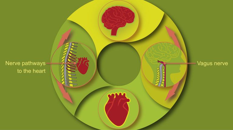
- People generally like to view the heart and brain as separate entities. Yet the brain – the center of thought – and our heart – the organ that pumps blood – have much in common and are in constant communication.
- If the heart muscle is weakened, the brain cells suffer as well.
- A Cluster of Excellence at the University Medical Center Göttingen is investigating the details of the interaction between the brain and the heart.
- Molecules, as well as entire networks of heart and nerve cells, are being studied at various scales.
- This is made possible by innovative imaging techniques such as optical nanoscopy.
- Researchers are investigating the precise interaction between brain and heart cells using mini-organs as models.
Heart muscle weakness is caused by a loss of heart muscle cells. It therefore makes sense to implant heart muscle cells into affected patients. A clinical trial is currently underway at the University Medical Center Göttingen to assess the safety and efficacy of a novel “heart patch” made from stem cells. It was also developed in Göttingen. The heart muscle tissue is applied to the diseased hearts of patients with heart failure to strengthen them.
The heart and brain are generally viewed as separate entities. But our brain and our heart are actually a true pair that still have a lot to say to each other, even after many years. And they stick together through thick and thin. If one is unwell, the other suffers too: About four million people in Germany suffer from heart failure – their heart muscle has been weakened by a heart attack or another condition and pumps less blood through the body. As a result, however, not only is physical performance limited, but often mental performance is as well: Heart problems can impair cognitive functions and increase the risk of dementia.
People with heart failure exhibit structural changes in the brain. For example, the Hippocampus – which is essential for learning and Memory – can shrink. Neuroscientist André Fischer from the University of Göttingen and the German Center for Neurodegenerative Diseases (DZNE) is interested in the underlying mechanisms. In a study, he and his colleagues examined mice that “only” suffered from heart failure. However, the affected mice subsequently performed worse on learning tasks than control mice. “We were able to show that, as a result of heart problems, these animals developed impaired Gene activity in the hippocampus,” says Fischer. “The DNA in their nerve cells was more tightly coiled, making it harder to read the genes.”
The reason for this tight coiling is chemical changes in the histones – the proteins responsible for packaging the DNA. As a result, various genes important for cognition and the hippocampus were less active than in healthy animals. Fischer and his colleagues had previously shown that people with Alzheimer’s disease exhibit such epigenetic changes in neurons and Glial cells “We assume that the reduced blood flow in the brain resulting from heart failure leads to such epigenetic changes.” The researchers also tested a drug in the mice that acts on histones and thus ultimately affects gene activity. “In fact, the animals’ learning performance improved again.”
Hippocampus
The hippocampus is the largest part of the archicortex and an area in the temporal lobe. It is also an important part of the limbic system. Functionally, it is involved in memory processes, but also in spatial orientation and learning. It comprises the subiculum, the dentate gyrus, and the Ammon's horn with its four fields CA1-CA4.
Changes in the structure of the hippocampus due to stress are associated with chronic pain. The hippocampus also plays an important role in the amplification of pain through anxiety.
Memory
Memory is a generic term for all types of information storage in the organism. In addition to pure retention, this also includes the absorption of information, its organization, and retrieval.
Gene
Information unit on DNA. Specialized enzymes translate the core component of a gene into ribonucleic acid (RNA). While some ribonucleic acids perform important functions in the cell themselves, others specify the order in which the cell should assemble individual amino acids into a specific protein. The gene thus provides the code for this protein. In addition, a gene also includes regulatory elements on the DNA that ensure that the gene is read exactly when the cell or organism actually needs its product.
Glial cells
Glia cells are the second largest group of cells in the brain after neurons. For a long time, they were considered inactive elements of the brain, referred to as "nerve cement." Today, we know that the different types of glia cells (astrocytes, oligodendrocytes, and microglia in the CNS; Schwann cells in the PNS) perform clearly defined tasks in the nervous system. For example, they respond to pathogens, play an important role in nourishing nerve cells, and insulate nerve fibers. They account for slightly more than 50 percent of the brain's cells, compared to neurons.
A revolution in imaging
The Cluster of Excellence “Multiscale Bioimaging: From Molecular Machines to Networks of Excitable Cells” focuses on the interaction between the brain and the heart.
The goal is to understand the function and structure of excitable heart and nerve cells. “And we want to investigate this using imaging and other methods at various levels,” says physician and neuroscientist Tobias Moser, director of the Institute for Auditory Neuroscience at the University Medical Center Göttingen. “From the smallest units – molecules – all the way up to networks of excitable cells.” In doing so, the researchers are studying molecular machines such as ion channels in the cell membrane, both individually and in their interactions with one another.
Research across different size scales has only recently become possible due to enormous technical advances. One such breakthrough is optical nanoscopy, which was developed in Göttingen by Nobel Prize laureate in Chemistry Stefan Hell. “We have thus succeeded, for example, in visualizing signal transmission at synapses and measuring the local calcium concentration at synapses,” says Moser. Until now, these signals could not be measured directly because their scale lies below the resolution limit of conventional light microscopy. Equally important to Moser and his colleagues is X-ray imaging, which is being developed in Göttingen in particular by experimental physicist Tim Salditt and his team. For Moser, it is the prime example of imaging across different size scales. It allows researchers to resolve very small objects but also to visualize entire organs. “X-ray microscopy enables us to visualize a large number of cells in heart and nerve tissue simultaneously, as well as at the cellular and network levels.”
The Göttingen researchers expect this research approach to yield insights that would not be possible by examining the heart or nerve tissue in isolation. And that makes sense. This is because heart and nerve cells contain the same ion channels, which are controlled by the voltage across cell membranes. This is the case, for example, in Hair cells in the inner ear.
Hearing in our nervous system occurs when these hair cells convert sound into neural signals that are transmitted to the brain. The hair cells have about a dozen synapses with the Auditory nerve fibers. In hair cells, the voltage-gated ion channels are ultimately the channels that provide calcium so that the important Neurotransmitter Glutamate can be released. “If such calcium channels fail due to a defect, the acoustic information cannot be transmitted from the ear to the brain, resulting in deafness,” says Moser. “In heart muscle cells, on the other hand, the channels are important for the propagation of electrical impulses.” If they fail due to a defect in the heart, a cardiac arrhythmia occurs.
Hair cells
Sensory cells in the inner ear located in the organ of Corti and the semicircular canals. The hair cells in the organ of Corti are responsible for transducing (converting) the vibrations into electrical potentials. Each of these sensory cells has hair-like protrusions of varying lengths, called stereocilia. These are interconnected. The movement of these stereocilia caused by the vibrations is the key to signal transduction in the hair cells.
Ear
auris
The ear is not only the organ of hearing, but also of balance. A distinction is made between the outer ear with the auricle and external auditory canal, the middle ear with the eardrum and ossicles, and the actual hearing and balance organ, the inner ear with the cochlea and semicircular canals.
Auditory nerve
nervus cochlearis
The hair cells of the organ of Corti stimulate neurons in the spiral ganglion, which is located in the cavity of the cochlea. Their axons form the auditory nerve, which transmits electrical impulses from the inner ear to the brain. Together with the vestibular nerve (nervus vestibularis), the auditory nerve forms the VIII cranial nerve.
Neurotransmitter
A neurotransmitter is a chemical messenger, an intermediary substance. It is released by the sender neuron at the sites of cell-cell communication and has an excitatory or inhibitory effect on the receiver neuron.
Glutamate
Glutamate is an amino acid and the most important excitatory neurotransmitter, which acts as a messenger substance in the transmission of information between neurons at their synapses.



Hidden similarities
There are other parallels between heart and nerve cells. In addition to the voltage-gated ion channels in the cell membrane, calcium-gated calcium channels are also found within heart and nerve cells.
Stephan Lehnart, a physician at the University Medical Center Göttingen, was able to demonstrate that when calcium flows into heart cells from outside, it triggers a veritable fireworks display. This is because the calcium-gated channels then open as well, releasing calcium into the interior of the cell. The heart muscle cell also needs this signal amplification so that the heart can contract. “Within the Cluster of Excellence, we are now also jointly investigating the role of calcium-gated calcium channels in hair cells,” says Moser.
Mini-brain as a test model
Scientists led by Maria-Patapia Zafeiriou at the University Medical Center Göttingen are investigating exactly how heart cells and nerve cells work together using so-called organoids. To do this, they develop organ-like structures in miniature format, such as “mini-brains,” which serve as models. To create them, human somatic cells are reprogrammed into a quasi-embryonic state. In this state, they can differentiate into almost any type of tissue, such as neural tissue. Such brain organoids have been around for some time. However, the researchers succeeded in creating fairly complex “mini-brains” measuring 5 to 7 millimeters – with interconnected excitatory and inhibitory neurons and supporting brain cells such as astrocytes. With the help of Zafeiriou’s “mini-brains,” it is possible, for example, to simulate and study the loss of learning ability and Memory in neurodegenerative diseases. In addition, drugs can be tested on the neural tissue.
But that’s not all. The researchers are particularly interested in the interaction between the heart and the brain. “We therefore merged our brain model with an already established heart muscle model,” says Zafeiriou. “After a few weeks in culture, we were able to see neural axons connecting with the heart muscle cells.” With the support of the Cluster of Excellence, the researchers then produced nerve cells that can be stimulated by light. In this model, stimulation at the interface between the brain and the heart increased the heart muscle’s heart rate. “This provides evidence of a functional interaction between certain nerve cells and heart muscle cells.”
So it’s becoming increasingly clear: the heart and the brain are a close-knit pair. In good times and bad, in sickness and in health.
excitatory
Exciting synapses are described as excitatory when they depolarize the subsequent cell membrane and can thus lead to the formation of an action potential. An excitatory effect is usually produced by an exciting transmitter (messenger substance), such as glutamate. The opposite is an inhibitory synapse.
Memory
Memory is a generic term for all types of information storage in the organism. In addition to pure retention, this also includes the absorption of information, its organization, and retrieval.