Development of a Brain
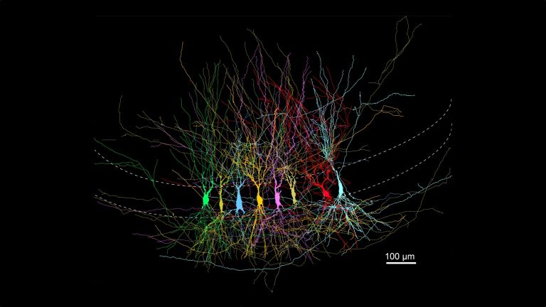
Our brain is the most complex structure we know: around 86 billion nerve cells, connected in a unique pattern by hundreds of trillions of synapses. It takes years to mature and is never truly finished.
Scientific support: Prof. Dr. Mark Hübener
Published: 10.02.2026
Difficulty: serious
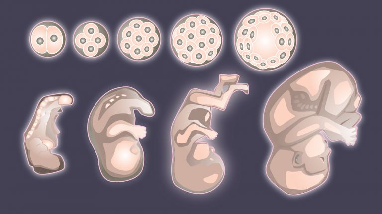
- Human development begins with numerous cell divisions. The fertilized egg cell develops into a morula, then a blastula. Within it, the cells that will form the embryo come together.
- Brain development begins around the 18th day with the closure of the neural tube.
- Later, the brain vesicles develop, followed by the rudiments of the large brain structures around the sixth week.
- The formation of the typical furrows of the Cerebrum begins around the 24th week and continues until around the first birthday.
- Growth factors and neighboring cells guide the neurons to their place in the brain.
- Neurons and synapses are initially formed in excessive numbers, with only the most stable ones remaining. This is how the brain adapts to its environment.
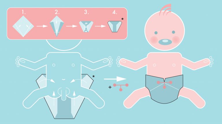
- The brain is by no means finished at birth; rather, enormous growth begins after birth.
- Radial Glial cells form the origin of many neurons.
Cerebrum
telencephalon
The cerebrum comprises the cerebral cortex (gray matter), the nerve fibers (white matter), and the basal ganglia. It is the largest part of the brain. The cortex can be divided into four cortical areas: the temporal lobe, frontal lobe, occipital lobe, and parietal lobe.
Its functions include the coordination of perception, motivation, learning, and thinking.
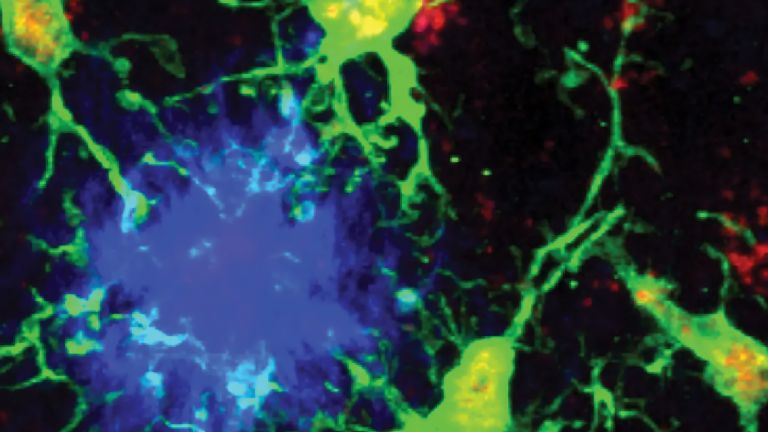
Glial cells
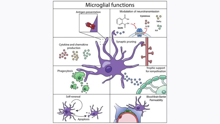
Glia cells are the second largest group of cells in the brain after neurons. For a long time, they were considered inactive elements of the brain, referred to as "nerve cement." Today, we know that the different types of glia cells (astrocytes, oligodendrocytes, and microglia in the CNS; Schwann cells in the PNS) perform clearly defined tasks in the nervous system. For example, they respond to pathogens, play an important role in nourishing nerve cells, and insulate nerve fibers. They account for slightly more than 50 percent of the brain's cells, compared to neurons.
A special type of glial cell, radial glial cells, plays an important role in brain development. Radial Glial cells arise from the epithelial cells of the neural tube at the beginning of neurogenesis. As "progenitor cells," they stand between stem cells and differentiated cells: studies show that they can produce some, but not all, cell types. Some of the radial glial cells produce other types of glial cells, including oligodendrocytes, which form the insulating sheaths of axons, and astrocytes, which initially act as guides and later, among other things, as nourishers of neurons. Another part, however, generates the neurons themselves during division. In the late stages of embryonic development and after birth, most radial glial cells have differentiated into other cell types. In a few parts of the brain, the remaining cells can continue to produce neurons into old age.
Glial cells
Glia cells are the second largest group of cells in the brain after neurons. For a long time, they were considered inactive elements of the brain, referred to as "nerve cement." Today, we know that the different types of glia cells (astrocytes, oligodendrocytes, and microglia in the CNS; Schwann cells in the PNS) perform clearly defined tasks in the nervous system. For example, they respond to pathogens, play an important role in nourishing nerve cells, and insulate nerve fibers. They account for slightly more than 50 percent of the brain's cells, compared to neurons.
Even the most complex structure in the body begins its development with the union of egg and sperm cells: approximately 24 hours after fertilization, the maternal and paternal chromosomes have combined to form the genetic makeup of the new individual. The first cell division begins. After 96 hours, the fertilized egg cell has developed into a sphere of about 30 cells that looks a little like a ripe mulberry, hence its name: morula. The cells of the morula are tiny because the first cells divide repeatedly into two halves without increasing in size. In the morula, the cells begin to differentiate into outer and inner cells. Three to four days after fertilization, fluid has collected in the center of the cell sphere – the morula has become a blastocyst. At one point in this hollow sphere, the innermost cells form a small cluster called the embryoblast. Only these cells develop into the embryo, while the rest become auxiliary organs such as the fetal parts of the placenta and the amniotic membranes.
While the embryo travels down the fallopian tube and the mother is still completely unaware of what is happening in her body, the division of labor in the developing human being progresses. The cells of the embryoblast fold into the three germ layers: endoderm, mesoderm, and ectoderm. The endoderm later develops into the internal organs, while the mesoderm develops into bones, muscles, and connective tissue. The ectoderm develops into the skin and, in a process called neurulation, the central nervous system, including the brain.
On the 18th day of the embryo's life, around the time when the mother realizes she may be pregnant, the first indentation forms in the ectoderm, which shortly thereafter constricts: the neural tube, the precursor to the Spinal cord Three protrusions, called brain vesicles, form at its front end. The embryo has now completed its journey and found a place in the uterine lining. It is now about two millimeters in size.
In the following days, the upper part of the neural tube with the brain vesicles bends slightly, and the first signs of the cerebral hemispheres become visible. Massive cell migration causes this area to enlarge significantly and become increasingly distinct from the spinal cord. Four weeks after fertilization, the Eye spots form and the heart begins to beat. After six weeks, the foundations for brain structures such as the Pons and cerebellum, thalamus, basal ganglia, and cerebral Cortex begin to develop. In the ninth week, tiny fingers and toes are already visible, and the spinal cord begins to control the first movements.
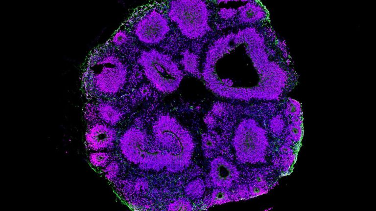
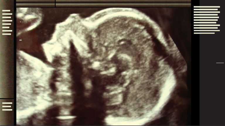
After three months, the embryo, now called a fetus, is twelve centimeters long and has well-developed structures in the Midbrain and hindbrain, but its cerebral cortex is still smooth and undifferentiated. It is not until around the 24th week that the first furrows typical of the human brain begin to form. This process continues after birth – until around the child's first birthday.
Spinal cord
medulla spinalis
The spinal cord is the part of the central nervous system located in the spine. It contains both the white matter of the nerve fibers and the gray matter of the cell nuclei. Simple reflexes such as the knee-jerk reflex are already processed here, as sensory and motor neurons are directly connected. The spinal cord is divided into the cervical, thoracic, lumbar, and sacral spinal cord.
Eye
bulbus oculi
The eye is the sensory organ responsible for perceiving light stimuli – electromagnetic radiation within a specific frequency range. The light visible to humans lies in the range between 380 and 780 nanometers.
Pons
pons
Area in the brain stem between the medulla oblongata and the mesencephalon. It acts as a switching station for many nerve pathways between the brain and spinal cord and contains numerous nuclei, including cranial nerves and those involved in controlling motor function in cooperation with the cerebellum.
Cortex
cortex cerebri
Cortex refers to a collection of neurons, typically in the form of a thin surface. However, it usually refers to the cerebral cortex, the outermost layer of the cerebrum. It is 2.5 mm to 5 mm thick and rich in nerve cells. The cerebral cortex is heavily folded, comparable to a handkerchief in a cup. This creates numerous convolutions (gyri), fissures (fissurae), and sulci. Unfolded, the surface area of the cortex is approximately 1,800cm².
Midbrain
mecencephalon
The midbrain is the uppermost section of the brain stem. Its regions are located around the aqueduct, a canal filled with cerebrospinal fluid. Prominent structures include the tectum, tegmentum, and substantia nigra.
Hiking trails through the brain
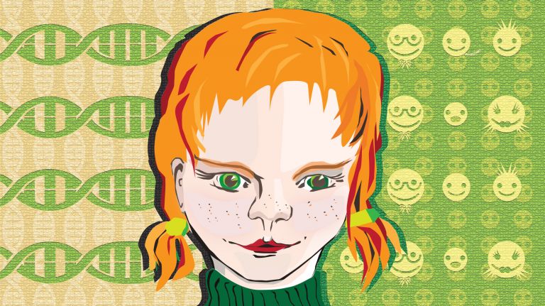
The structure of the human brain is only genetically determined in broad terms. Its fine structure is the result of a complex organizational process in which environmental factors also play a role. These include the mother's diet and any illnesses or exposure to toxins. ▸ Like mother, like Child
The young neurons develop from stem cells in a tissue layer of the neural tube. To get a rough idea of this process, you can divide the number of neurons in the brain by the months of pregnancy: this gives an average value of 250,000 new neurons per minute. From there, they migrate to their destinations in the brain and begin to specialize for their tasks during this migration: for example, into photoreceptor cells or olfactory cells. Their tasks depend on when they were created and on chemical factors in their environment. First, the inner layers of the Cerebrum develop, and the younger cells migrate past the older ones to form the outer layer. In doing so, they use the radial glial cells, whose long processes grow outward across the layers of the brain, as a kind of railing to hold onto (see info box).
Once a Neuron has arrived at its destination, it must connect to its target region. If a neuron is located in the Retina of the eye, for example, it must dock onto the visual center in the thalamus. To do this, it extends an "arm," a neurite, at the tip of which is a growth cone ▸ The Neuron: Form and Function (interactive). It paves the way for the neurite, similar to an axon, through the dense tissue, sometimes even reaching the other half of the brain. Where this growth cone grows is determined, on the one hand, by attractive and repulsive substances on the surfaces of the surrounding cells. "This allows proper roads or channels to form, which the neurites use for orientation," explains Paul G. Layer, professor emeritus of developmental biology and neurogenetics at Darmstadt Technical University. On the other hand, growth factors influence where the neurite extends. These are small proteins that are emitted by the target regions of the neurites and can be detected by the growth cone with receptors on its numerous tentacles. "Unlike the substances on the cell surfaces, growth factors can act over certain distances," explains Layer. The neurite then grows to where the concentration of the growth factor is highest.
Cerebrum
telencephalon
The cerebrum comprises the cerebral cortex (gray matter), the nerve fibers (white matter), and the basal ganglia. It is the largest part of the brain. The cortex can be divided into four cortical areas: the temporal lobe, frontal lobe, occipital lobe, and parietal lobe.
Its functions include the coordination of perception, motivation, learning, and thinking.
Neuron
A neuron is a specialized cell in the nervous system that is responsible for processing and transmitting information. It receives signals via its dendrites and transmits them via its axon. Transmission occurs electrically within the neuron and, between neurons, usually chemically via synapses.
Retina
The retina is the inner layer of the eye covered with pigment epithelium. The retina is characterized by an inverse (reversed) arrangement: light must first pass through several layers before it hits the photoreceptors (cones and rods). The signals from the photoreceptors are transmitted via the optic nerve to the processing areas of the brain. The reason for the inverse arrangement is the evolutionary development of the retina, which is a protrusion of the brain.
The retina is approximately 0.2 to 0.5 mm thick.


Those who are not needed die
Once the growth cone has reached its destination, the Neuron is still far from finished. "First, the cell Nucleus must be informed that the neurite has arrived," says Layer: "To do this, the growth factor must be transported backwards through the neurite into the cell body. Once there, it triggers a signaling cascade that activates genes in the cell nucleus that are necessary for the neuron's survival. If this signal is not received because the neurite has not found its target, the neuron undergoes apoptosis, which means it commits suicide." The growth factor is therefore more of a survival factor.
In fact, the mass production of neurons in the embryo's brain leads to there being far too many neurons at times. They compete with each other: only those with the most stable connections remain. Up to 80 percent of neurons are broken down again.
Most of the successful axons are later enveloped by extensions of the Oligodendrocytes. This process, known as myelination, begins in the oldest structure of the brain, the Brain stem ▸ The Brain Stem, and continues into the youngest ones. They isolate the axons from the processes in their environment and enable the electrical impulses with which the cells communicate to be transmitted up to a hundred times faster ▸ Nerve Cells in Conversation. Myelination continues into adulthood in the Frontal lobe of the Cerebrum. ▸ The Frontal Lobe
Neuron
A neuron is a specialized cell in the nervous system that is responsible for processing and transmitting information. It receives signals via its dendrites and transmits them via its axon. Transmission occurs electrically within the neuron and, between neurons, usually chemically via synapses.
Nucleus
In cell biology, the nucleus in a cell is the cell nucleus, which contains the chromosomes, among other things. In neuroanatomy, the nucleus in the nervous system refers to a collection of cell bodies – known as gray matter in the central nervous system and ganglia in the peripheral nervous system.
Oligodendrocytes
Cells of the central nervous system that form the myelin sheath around nerve cells, thereby increasing their conduction velocity. They belong to the glial cells.
Brain stem
truncus cerebri
The "trunk" of the brain, to which all other brain structures are "attached," so to speak. From bottom to top, it comprises the medulla oblongata, the pons, and the mesencephalon. It transitions into the spinal cord below. It is a center for vital functions such as breathing and heartbeat and contains ascending and descending pathways between the cerebrum, cerebellum, and spinal cord.
frontal
An anatomical position designation – frontal means "towards the forehead," i.e., at the front.
Frontal lobe
Lobus frontalis
The frontal cortex is the largest of the four lobes of the cerebral cortex and its functions are correspondingly comprehensive. The front area, known as the prefrontal cortex, is responsible for complex action planning (known as executive functions), which also shapes our personality. Its development (myelination) takes up to 30 years and even then is not yet complete. Other important components of the frontal cortex are Broca's area, which controls our ability to express ourselves linguistically, and the primary motor cortex, which sends movement impulses throughout the body.
Cerebrum
telencephalon
The cerebrum comprises the cerebral cortex (gray matter), the nerve fibers (white matter), and the basal ganglia. It is the largest part of the brain. The cortex can be divided into four cortical areas: the temporal lobe, frontal lobe, occipital lobe, and parietal lobe.
Its functions include the coordination of perception, motivation, learning, and thinking.
Finally outside
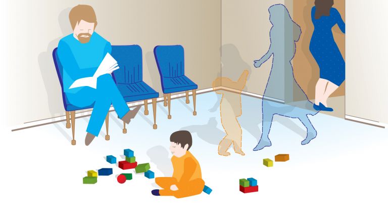
The brain is by no means fully developed at birth. Birth is rather the starting point for an enormous growth spurt: in the first year of life, the brain grows threefold and the head reaches three-quarters of its adult size. This growth spurt is partly due to the formation of new neurons and partly to the fact that the nerve fibers become thicker through myelination and the neurons establish numerous synapses with their neighbors. This formation of synapses initially occurs rapidly and indiscriminately, as if one were trying to get to know everyone at a party through "speed dating." At the age of three, children have twice as many synapses as adults. At the party, you eventually have to decide who you actually want to talk to. The neurons experience something similar: only the most active synapses remain, while the others are broken down. This process is called synaptic pruning and continues in vertebrates until puberty. During childhood, it can cost us billions of synapses every day. That sounds alarming, but it actually leads to more efficient cognitive processes.
This slow development of the brain, heavily influenced by the experiences a child has in its environment, makes humans so adaptable, so intelligent, but also so impressionable.
Further reading
- Dan H. Sanes, Thomas A. Reh, William A. Harris: Development oft he Nervous System. Elsevier Publisher 2012.
- Lise Eliot: What's Going on in There?: How the Brain and Mind Develop in the First Five Years of Life. 1999
Originally published on March 17, 2016
Last updated on February 20, 2026