Children in the Lab
Babies and children are particularly valuable for brain research, as many key developmental processes can only be studied in the early stages of life. At the same time, research involving minors poses significant challenges – both practical and ethical.
Scientific support: Prof. Dr. Claudia Buß
Published: 18.03.2026
Difficulty: intermediate
- In basic research and clinical trials, babies and children are highly sought-after participants.
- To turn them into committed volunteers, researchers design their studies to be child-friendly: entertaining and engaging.
- Various methods such as EEG or near-infrared spectroscopy are used in research with children. Magnetic resonance imaging (MRI) scans are a challenge: the little ones must not move and need to be psychologically prepared for the confined space and noise of the machine.
- Ethicists place particular emphasis on ensuring that risks and stress for the children are minimal; after all, the children derive no direct benefit from basic research.
EEG
An electroencephalogram, or EEG for short, is a recording of the brain's electrical activity (brain waves). Brain waves are measured on the surface of the head or using electrodes implanted in the brain itself. The time resolution is in the millisecond range, but the spatial resolution is very poor. The discoverer of electrical brain waves and EEG is the neurologist Hans Berger (1873−1941) from Jena.
Magnetic resonance imaging
Magnetic resonance imaging scanner
A device used by medical professionals for magnetic resonance imaging (MRI). MRI is an imaging technique used to diagnose malformations in various tissues or organs of the body. This method is particularly effective for imaging parts of the body that contain a lot of water. Patients are placed in a tube (scanner) and exposed to a strong magnetic field. However, they are not exposed to X-rays or other forms of ionizing radiation.
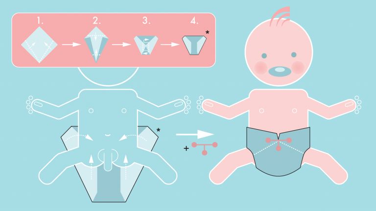
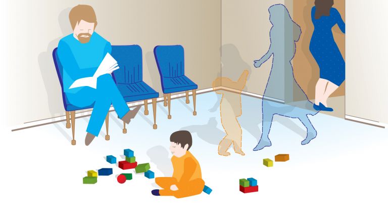
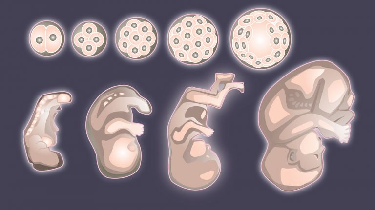
Many research questions – such as those regarding the origins of mental illnesses – must be addressed during phases when the brain is still particularly sensitive to environmental influences. Since the brain is highly plastic, especially in early childhood, this developmental phase is of particular interest to science. Specialized baby labs, where babies and toddlers are studied, are tailored entirely to the specific needs of their young participants. Here, you’ll find bottle warmers in the kitchens, changing rooms, and toys in the waiting area. The labs themselves, however, are usually kept deliberately unadorned. Nothing for a child’s gaze to linger on – after all, little ones are easily distracted. Hungry or tired babies also make for unwilling participants. So the experiments, meals, and short naps must be carefully coordinated. During the trials, researchers rely on photos and videos on computer screens; above all, these must be colorful and interesting. If none of that helps, they sometimes play music to keep the little ones engaged. Or they use “stimulating” sounds to bring the little ones back to their task in front of a screen. In addition to measuring brain waves via electrodes, an important research method is the so-called gaze duration measurement or gaze preference method. In this method, researchers observe how long the gaze of babies and toddlers lingers on an image or video element. On average across all participants, a difference can often be detected: do the babies, on average, look at one image or another for longer? The researchers then infer preferences or irritations from these differences.
A roar fills the spaceship. Landscapes and places pass before Neo’s eyes. The voice of the ground station sounds intermittently through the speakers.
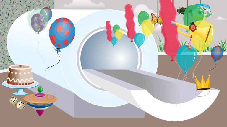
This isn’t a scene from a science fiction movie. The action takes place right here on Earth at the Max Planck Institute for Human Development in Berlin. Neo is a participant in a neuroscientific study on Memory – and he’s only six years old. The experiment is tailored to young volunteers like him and is therefore packaged as a story: The children are astronauts, the MRI scanner is a spaceship, and the lab is the ground station.
Whether researchers want to study brain development in basic research or the effects of treatments in clinical trials, children and babies are highly sought-after participants in neuroscience (see info box). But brain research involving minors is anything but child’s play. Even though children’s games are exactly what researchers often offer their young subjects to keep them entertained during the experiments.
Memory
Memory is a generic term for all types of information storage in the organism. In addition to pure retention, this also includes the absorption of information, its organization, and retrieval.
Oskar the stuffed animal explains the procedures
An hour and a half beforehand: Researchers at the Max Planck Institute in Berlin are carefully preparing Neo for the scans in the Magnetic resonance imaging (MRI) machine. Among other things, functional measurements are to be taken. Even small movements of the head or neck can compromise the quality of the fMRI images. Using the stuffed animal Oskar, a research assistant explains to Neo why it’s so important to stay still. “It’s like taking a regular photo of you,” she says. “Here, he moved.” Oskar appears blurry in an image on the computer monitor.
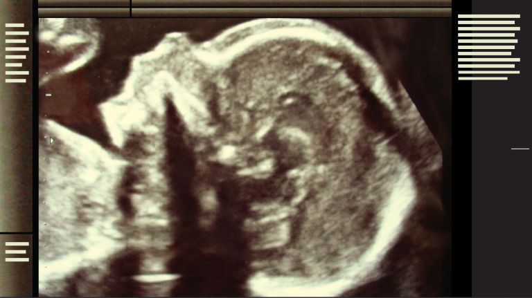
Lying still in a scanner is no easy task for the young participants; after all, depending on the examination, the scans can take up to an hour. The fact that MRI is so sensitive to movement is also why brain scans of babies and toddlers are rarely performed in basic research. There are only two options: sedating the little ones, which is ethically questionable – or hoping that the sandman comes on his own. Psychologist Alice Graham and her colleagues at the University of Oregon did the latter in a study published in 2013. They found that even babies’ brains register the emotional tone of voices – even when the little ones are blissfully slumbering in the scanner, as in the study.
Of course, many things cannot be studied this way, such as how the brain reacts to visual stimuli. That is why researchers rely on other methods, especially when working with babies and toddlers. With electroencephalography (EEG) using electrodes on the scalp, a child can sit comfortably on their mother’s lap and view stimuli presented on a screen. However, this method offers poor spatial resolution and measures only processes on the brain’s surface.
Consequently, neuroscientists are increasingly using near-infrared spectroscopy (NIRS) to send long-wavelength infrared light through a child’s skull. The reflected light allows them to estimate neural activity. Compared to EEG, NIRS allows for a deeper look into the brain, and the young subjects can move freely during the measurements without interfering with the signal. But even NIRS cannot penetrate deeper regions of the brain such as the Hippocampus and the Amygdala. That is reserved for MRI.
Magnetic resonance imaging
Magnetic resonance imaging scanner
A device used by medical professionals for magnetic resonance imaging (MRI). MRI is an imaging technique used to diagnose malformations in various tissues or organs of the body. This method is particularly effective for imaging parts of the body that contain a lot of water. Patients are placed in a tube (scanner) and exposed to a strong magnetic field. However, they are not exposed to X-rays or other forms of ionizing radiation.
EEG
An electroencephalogram, or EEG for short, is a recording of the brain's electrical activity (brain waves). Brain waves are measured on the surface of the head or using electrodes implanted in the brain itself. The time resolution is in the millisecond range, but the spatial resolution is very poor. The discoverer of electrical brain waves and EEG is the neurologist Hans Berger (1873−1941) from Jena.
Hippocampus
The hippocampus is the largest part of the archicortex and an area in the temporal lobe. It is also an important part of the limbic system. Functionally, it is involved in memory processes, but also in spatial orientation and learning. It comprises the subiculum, the dentate gyrus, and the Ammon's horn with its four fields CA1-CA4.
Changes in the structure of the hippocampus due to stress are associated with chronic pain. The hippocampus also plays an important role in the amplification of pain through anxiety.
Amygdala
corpus amygdaloideum
An important core area in the temporal lobe that is associated with emotions: it evaluates the emotional content of a situation and reacts particularly to threats. In this context, it is also activated by pain stimuli and plays an important role in the emotional evaluation of sensory stimuli. Inaddition, it is involved in linking emotions with memories, emotional learning ability, and social behavior. The amygdala is part of the limbic system.
Psychologically stressful?
In Berlin, researchers are now familiarizing Neo with the MRI scanner. Some participants find it distressing to be confined in the narrow tube; some even have claustrophobia or experience a panic attack. Others are bothered by the noise – the loud knocking sounds of the scanner. At the Max Planck Institute, staff therefore ask parents in advance whether their child shows signs of agoraphobia or claustrophobia. If so, the children cannot participate in the study. To psychologically prepare the children for the scanner, the researchers use an MRI simulator that looks strikingly similar to the real device. “And just in case, there’s also an emergency button,” says Attila Keresztes, a cognitive neuroscientist and one of the researchers involved in the study. “The children can press this button if they feel uncomfortable. Then the test is paused.”
In the scientific literature, it has so far been debated whether MRI scans are more stressful for children than for adults. For this reason, researchers led by psychologist Charlotte Jaite from Charité in Berlin have just conducted a corresponding study. The scientists examined whether and to what extent MRI examinations trigger negative Emotions in children and adolescents. According to Section 41, Paragraph 2 of the German Medicines Act, which regulates, among other things, the testing of drugs, research must not impose more than “minimal stress” on minors. Jaite’s team therefore compared the effects of MRI scans with those of EEG examinations. After all, the latter are currently considered “minimal burden.” “Based on our observations to date during data collection, pediatric and adolescent patients do not appear to exhibit higher anxiety levels before and during an MRI scan than children and adolescents undergoing EEG examinations.” The researchers wrote this in 2013 in a summary of their study design.
Emotions
Neuroscientists understand "emotions" to be complex response patterns that include experiential, physiological, and behavioral components. They arise in response to personally relevant or significant events and generate a willingness to act, through which the individual attempts to deal with the situation. Emotions typically occur with subjective experience (feeling), but differ from pure feeling in that they involve conscious or implicit engagement with the environment. Emotions arise in the limbic system, among other places, which is a phylogenetically ancient part of the brain. Psychologist Paul Ekman has defined six cross-cultural basic emotions that are reflected in characteristic facial expressions: joy, anger, fear, surprise, sadness, and disgust.
EEG
An electroencephalogram, or EEG for short, is a recording of the brain's electrical activity (brain waves). Brain waves are measured on the surface of the head or using electrodes implanted in the brain itself. The time resolution is in the millisecond range, but the spatial resolution is very poor. The discoverer of electrical brain waves and EEG is the neurologist Hans Berger (1873−1941) from Jena.


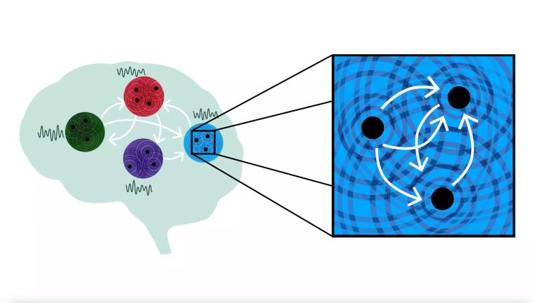
Pattern separation in the brain
After Neo has spent some time getting used to the simulator, it’s now time for the real scanner. “Hello Captain Neo, this is ground control,” comes the voice from the speaker. “Are we launching your rocket now?” “Yes,” replies the brave astronaut. In keeping with the astronaut story, he is supposed to press a button every time he spots an alien in the images showing him various landscapes and locations. Sometimes the scenes repeat themselves; other times, they are only very similar. “The study focuses on how so-called pattern separation in the brain develops over the course of a lifetime,” explains Attila Keresztes. “This function of Memory allows us to store two very similar experiences differently.” The Berlin researchers suspect that a subregion of the seahorse-shaped Hippocampus plays an important role in such interwoven memories.
To draw out the secrets from Neo’s head, he is now surrounded by a strong magnetic field and radio waves inside the scanner. Unlike computed tomography, for example, MRI does not produce ionizing X-rays. “Physically speaking, the MRI scan has no harmful effects,” says Attila Keresztes. Studies confirm this. Three decades of MRI use in humans have shown no acute effects or long-term biological consequences. This was reported by researchers led by biostatistician Mekibib Altaye of the University of Cincinnati College of Medicine in a study from 2014. Most regulatory bodies therefore classify MRI scans as posing a minimal risk. In their own long-term study, the researchers also found no statistically significant evidence that the mental or physical development of child participants had suffered over a ten-year period due to annual MRI scans. However, this was only a small study with few participants, and the researchers used only weight, height, and body mass index as physical markers.
Memory
Memory is a generic term for all types of information storage in the organism. In addition to pure retention, this also includes the absorption of information, its organization, and retrieval.
Hippocampus
The hippocampus is the largest part of the archicortex and an area in the temporal lobe. It is also an important part of the limbic system. Functionally, it is involved in memory processes, but also in spatial orientation and learning. It comprises the subiculum, the dentate gyrus, and the Ammon's horn with its four fields CA1-CA4.
Changes in the structure of the hippocampus due to stress are associated with chronic pain. The hippocampus also plays an important role in the amplification of pain through anxiety.
Ethical Aspects of Research Involving Children
Either way, ethical concerns remain. “Studies involving minors present a tricky moral problem,” says philosopher Bert Heinrichs of the Institute for Science and Ethics at the University of Bonn. “Clinical trials are primarily about scientific findings and, at most, secondarily about benefits for the participants.” This applies all the more to basic research. “With adults, this is generally not a problem; they can give their consent and they know what they’re getting into.” But depending on their age, children are either not capable of giving consent at all or only to a limited extent. “On the other hand, without any research on minors, we wouldn’t have medications tailored for children,” says Heinrichs.
In Germany, there is no specific law regulating research on humans. There are only the Medicines Act and the Medical Devices Act, each of which regulates specific aspects. “Otherwise, we have general laws such as the Civil Code,” adds Heinrichs. This stipulates that parents are obligated to act in the best interests of the child when making decisions that affect the child. “In my view, therefore, both the risk and the burden must not exceed a minimal level.” Yet the problem remains: which examinations meet these criteria?
Neo seems to have survived his long wait in his spaceship unscathed. “Good,” he replies somewhat shyly when asked how it went. And then he breaks into a broad smile, a typical child’s smile.
Further reading
- Holland, S.K. et al.: Data on the safety of repeated MRI in healthy children. Neuroimage Clin. 2014 Feb 9;4:526–3, (abstract).
Originally published on March 18, 2016
Last updated on March 18, 2026