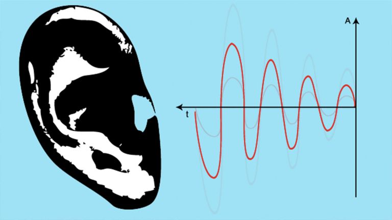
Hearing: Structure of the Organ of Corti
Published: 23.10.2025
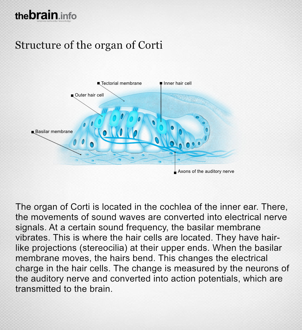
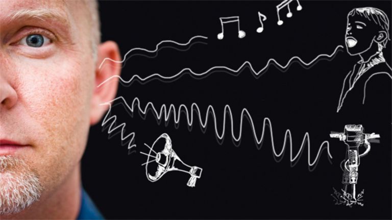
The Hair cells are important in the organ of Corti. They change their electrical charge as soon as the Basilar membrane vibrates. This creates a hearing impression in the brain.
You can read more about this in our article ▸ From Wiggling to the wonderful Variety of Sounds.
Hair cells
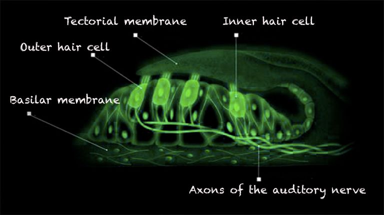
Sensory cells in the inner Ear located in the organ of Corti and the Semicircular canals The Hair cells in the organ of Corti are responsible for transducing (converting) the vibrations into electrical potentials. Each of these sensory cells has hair-like protrusions of varying lengths, called stereocilia. These are interconnected. The movement of these stereocilia caused by the vibrations is the key to signal transduction in the hair cells.
Basilar membrane
The Basilar membrane runs through the Cochlea for a length of approximately 34 mm. It is stretched like the string of a violin, narrow and stiff at the base and wider and more flexible at the apex. Incoming sound frequencies cause it to vibrate. This movement is picked up by the hair cells in the organ of Corti and converted into nerve impulses.
Ear
auris
The ear is not only the organ of hearing, but also of balance. A distinction is made between the outer ear with the auricle and external auditory canal, the middle ear with the eardrum and ossicles, and the actual hearing and balance organ, the inner ear with the cochlea and semicircular canals.
Semicircular canals
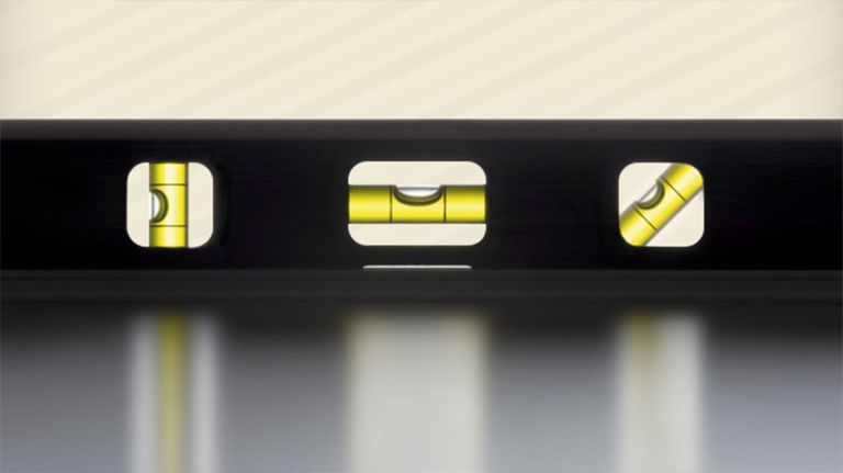
The three semicircular canals per ear are interconnected, fluid-filled tubes that are positioned almost at right angles to each other and belong to the balance organ in the inner ear (vestibular apparatus). They serve to register angular accelerations, i.e., rotational movements of the head.
Hair cells
Sensory cells in the inner ear located in the organ of Corti and the semicircular canals. The hair cells in the organ of Corti are responsible for transducing (converting) the vibrations into electrical potentials. Each of these sensory cells has hair-like protrusions of varying lengths, called stereocilia. These are interconnected. The movement of these stereocilia caused by the vibrations is the key to signal transduction in the hair cells.
Basilar membrane
The basilar membrane runs through the cochlea for a length of approximately 34 mm. It is stretched like the string of a violin, narrow and stiff at the base and wider and more flexible at the apex. Incoming sound frequencies cause it to vibrate. This movement is picked up by the hair cells in the organ of Corti and converted into nerve impulses.
Cochlea
The cochlea is the part of the inner ear that contains the organ of Corti, which is responsible for converting acoustic signals into nerve impulses.