Protective Brain Immune Cell State Discovered
Discovery points to a potential new therapeutic pathway for Alzheimer’s disease
Published: 05.11.2025
To the point:
- Protective Microglia identified: In Alzheimer’s disease, microglia can become “inflamed,” contributing to neuronal damage. Researchers have now discovered that this inflammatory activity can be suppressed by a small subpopulation of neuroprotective microglia.
- Regulatory functions: Specialized cells express a distinctive set of immunoregulatory lymphoid Receptor proteins, including CD28. Much like suppressor T cells that control autoimmunity in the peripheral immune system, this CD28-expressing microglial population can dampen brain-wide microglial inflammation and slow the buildup of amyloid Plaques - a hallmark of Alzheimer’s disease.
- Therapeutic promise: Discovery reveals a new immunoregulatory pathway that could be harnessed to develop therapies enhancing microglial protective functions to slow or prevent Alzheimer’s disease progression.
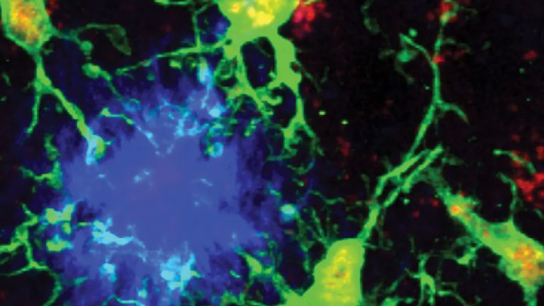
In Alzheimer’s disease—the leading cause of dementia—microglia, the brain’s immune defenders, can act as both protectors and aggressors, shaping how the disease progresses. Researchers at the Max Planck Institute for Biology of Ageing in Cologne and the Icahn School of Medicine at Mount Sinai in New York, in close collaboration with The Rockefeller University, The City University of New York and multiple international partners, have identified a distinct population of neuroprotective microglia, that may point to a new therapeutic approach for Alzheimer’s disease. In a study published in Nature, the team reports that microglia with reduced expression of the transcription factor PU.1 and co-expression of the lymphoid-like receptor CD28 act to limit neuroinflammation and to slow amyloid-plaque build-up and neurotoxic Tau protein spreading in the brain, the major hallmarks of Alzheimer’s pathology.
Using Alzheimer’s mouse models, human cells, and human brain tissue, the researchers demonstrated that lowering PU.1 promotes the expression of lymphoid immunoregulatory receptor proteins on microglia. Despite being present in small numbers, these neuroprotective microglia exert a brain-wide suppressive impact on inflammation and protect cognitive function and survival in mice. Deleting CD28 from this small subset of microglia amplified inflammation and accelerated plaque growth, highlighting CD28’s key role in protective microglial activity.
Microglia are not simply destructive responders in Alzheimer’s disease - they can become the brain’s protectors. Anne Schaefer
“Microglia are not simply destructive responders in Alzheimer’s disease— they can become the brain’s protectors,” said Anne Schaefer, the senior author of the paper and leader of the project. “This finding extends our earlier observations on the remarkable Plasticity of microglia states and their important roles in diverse brain functions. It also underscores the vital importance of international collaboration in advancing scientific progress.”
“It is remarkable to see that molecules long known to immunologists for their roles in B and T lymphocytes also regulate microglial activity,” added Alexander Tarakhovsky. “This discovery comes at a time when regulatory T cells have achieved major recognition as master regulators of immunity, highlighting a shared logic of immune regulation across cell types. It also paves the way for immunotherapeutic strategies for Alzheimer’s disease.”
The study builds on pioneering genetic work by Alison Goate, a senior co-author of the study, who identified a common variant in SPI1—the Gene encoding PU.1—as being associated with reduced Alzheimer’s risk. “These results provide a mechanistic explanation for why lower PU.1 levels are linked to reduced Alzheimer’s risk,” said Goate.
The discovery of the PU.1–CD28 axis establishes a molecular framework for understanding protective microglial states and highlights the potential of microglia-targeted immunotherapies to modify the course of Alzheimer’s disease.
Microglia
The smallest type of glial cell is part of the cellular immune system and is responsible, among other things, for removing dead neurons. Microglia can move in an amoeba-like manner.
Receptor
A receptor is a protein, usually located in the cell membrane or inside the cell, that recognizes a specific external signal (e.g., a neurotransmitter, hormone, or other ligand) and causes the cell to trigger a defined response. Depending on the type of receptor, this response can be excitatory, inhibitory, or modulatory.
Plaques
Senile plaques
Senile plaques accumulate in the gray matter of the brain when a protein – known as amyloid precursor protein – is not broken down correctly. Inflammation and disorders of fat or sugar metabolism can promote plaque formation. On average, the deposits reach a diameter of 50 micrometers. The appearance of plaques is one of several anatomical changes in the brain that pathologists can use to diagnose Alzheimer's disease after death.
Tau protein
Tau proteins are particularly prevalent in the central nervous system. Their function is to stabilize microtubules – the structures that give cells their shape and support. Under certain circumstances, enzymes attach too many phosphate groups to tau proteins. As a result, the proteins are no longer broken down properly and form toxic aggregates within the neurons. Alongside senile plaques, aggregated tau proteins are considered a classic hallmark of Alzheimer's disease.
Plasticity
Neuroplasticity
The term neuroplasticity describes the ability of synapses, nerve cells, and entire areas of the brain to change structurally and functionally depending on the degree to which they are used. Synaptic plasticity refers to the adaptation of the signal transmission strength of synapses to the frequency and intensity of incoming stimuli, for example in the form of long-term potentiation or depression. In addition, the size, interconnection, and activity patterns of different areas of the brain also change depending on their use. This phenomenon is referred to as cortical plasticity when it specifically affects the cortex.
Gene
Information unit on DNA. Specialized enzymes translate the core component of a gene into ribonucleic acid (RNA). While some ribonucleic acids perform important functions in the cell themselves, others specify the order in which the cell should assemble individual amino acids into a specific protein. The gene thus provides the code for this protein. In addition, a gene also includes regulatory elements on the DNA that ensure that the gene is read exactly when the cell or organism actually needs its product.
