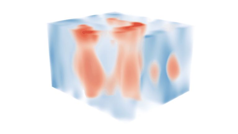
The Cortex

Even non-anatomists have this characteristic image of the brain in their minds: a helmet-shaped structure with a surface covered in convolutions and furrows. This outermost part of the brain – well protected by the skull and the meninges underneath – is the cerebral cortex. It usually is referred to simply as the cortex, the Latin word for “bark.”
Scientific support: Prof. Dr. Karl Zilles, Prof. Dr. Andreas Vlachos
Published: 20.09.2025
Difficulty: intermediate
The cerebral Cortex covers almost the entire brain visible from the outside. It is heavily folded and crisscrossed by numerous furrows, creating distinct areas. Each half of the Cerebrum (Hemisphere) is divided into four lobes that are visible from the outside: the frontal lobe, parietal lobe, temporal lobe, and Occipital lobe In addition, there is the insular lobe (lobus insularis), which is hidden deep in the lateral cerebral sulcus and is not visible from the outside. About 90 percent of the cortex consists of the evolutionarily young neocortex, which is composed of six cell layers throughout. The older parts in terms of evolutionary development – the Paleocortex and Archicortex – differ in their “simpler” cellular structure.
The cortex is the biological basis of all our higher mental abilities.
Cortex
cortex cerebri
Cortex refers to a collection of neurons, typically in the form of a thin surface. However, it usually refers to the cerebral cortex, the outermost layer of the cerebrum. It is 2.5 mm to 5 mm thick and rich in nerve cells. The cerebral cortex is heavily folded, comparable to a handkerchief in a cup. This creates numerous convolutions (gyri), fissures (fissurae), and sulci. Unfolded, the surface area of the cortex is approximately 1,800cm².
Cerebrum
telencephalon
The cerebrum comprises the cerebral cortex (gray matter), the nerve fibers (white matter), and the basal ganglia. It is the largest part of the brain. The cortex can be divided into four cortical areas: the temporal lobe, frontal lobe, occipital lobe, and parietal lobe.
Its functions include the coordination of perception, motivation, learning, and thinking.
Hemisphere
The cerebrum and cerebellum each consist of two halves – the right and left hemispheres. In the cerebrum, they are connected by three pathways (commissures). The largest commissure is the corpus callosum.
frontal
An anatomical position designation – frontal means "towards the forehead," i.e., at the front.
Occipital lobe
lobus occipitalis
One of the four large lobes of the cerebral cortex. The occipital lobe lies above the cerebellum. It borders the parietal and temporal lobes at the front. The calcarine sulcus divides the occipital lobe into an upper and lower half, the cuneus and the lingual gyrus. Functionally, this area of the brain is responsible for the central processing of visual information – both the primary and secondary visual cortex are located in the occipital lobe.
lateral
A positional term – lateral means "towards the side." In relation to the nervous system, it refers to a direction at right angles to the neural axis, i.e., to the right or left.
Paleocortex
The paleocortex is a phylogenetically very old part of the telencephalon, which together with the olfactory bulb forms the olfactory brain. The paleocortex differs from the isocortex in that it does not have a six-layer structure.
Archicortex
An ancient structure of the cerebrum in terms of evolutionary development, which, in contrast to the isocortex (also called the neocortex), has a three-layer structure. The archicortex mainly comprises the hippocampal structures.
The cerebrum, with its two hemispheres and the corpus callosum connecting them, is the youngest and largest part of the brain in terms of evolutionary development. It accounts for about 85 percent of the total brain mass. If we remove the cerebral medulla – consisting mainly of nerve fibers and the Basal ganglia (nuclei basales) embedded in it – we are left with the cerebral cortex, a two- to five-millimeter-thick layer known as gray matter. It is rich in nerve cell bodies, which give it a reddish-brown to gray color. Estimates suggest that there are around 17 billion nerve cells (neurons) in the human cerebral Cortex. Individual differences between women and men are primarily related to the larger average brain and body size of men – they do not allow any conclusions to be drawn about mental abilities.
So, size does not matter. But mental abilities do: in the cortex, signals from the sensory organs and upstream brain regions are combined to form a coherent impression of the environment. It can also store information, thus forming the biological basis of our Memory. Thinking, goal-oriented action, feelings – in short, all higher mental and psychological functions of humans – are not possible without the cerebral cortex.
Basal ganglia
Nuclei basales
The basal ganglia are a group of subcortical nuclei (located beneath the cerebral cortex) in the telencephalon. The basal ganglia include the globus pallidus and the striatum, and, depending on the author, other structures such as the substantia nigra and the subthalamic nucleus. The basal ganglia are primarily associated with voluntary motor function, but they also influence motivation, learning, and emotion.
Cortex
cortex cerebri
Cortex refers to a collection of neurons, typically in the form of a thin surface. However, it usually refers to the cerebral cortex, the outermost layer of the cerebrum. It is 2.5 mm to 5 mm thick and rich in nerve cells. The cerebral cortex is heavily folded, comparable to a handkerchief in a cup. This creates numerous convolutions (gyri), fissures (fissurae), and sulci. Unfolded, the surface area of the cortex is approximately 1,800cm².
Memory
Memory is a generic term for all types of information storage in the organism. In addition to pure retention, this also includes the absorption of information, its organization, and retrieval.
The evolution of intelligence and consciousness
The typical structure of the Cortex has slowly developed into its present form over the course of mammalian evolution. First, the part responsible for smell Perception developed – it is therefore called the paleocortex, or old cortex. The so-called Archicortex also developed very early on. It is often counted as part of the Limbic system and, in humans, comprises parts of the cerebral cortex that are responsible for emotional responses, behavior for species preservation, and reproduction. In addition, there is the hippocampus, which is of central importance for Memory and spatial orientation.
However, these “old” areas only make up about one-tenth of the cerebral cortex. The remaining 90 percent form the Neocortex. With the increasing development and refinement of the senses in mammals – which include not only the eyes, ears, and Taste organs, but also the sensory receptors in the skin, mucous membranes, and muscles, as well as the Retina and inner Ear with the hearing and balance systems – the neocortex became increasingly complex as well. In addition to motor areas for controlling specific movements, it primarily comprises large sections of the so-called association cortex.
In the association cortex, information from the many sensory systems is combined to form a comprehensive picture of the world; this is also where our Attention and activity are regulated. The Association cortex not only processes sensory impressions that enter the brain from outside, but also incorporates internal processes such as memories, expectations, and thoughts. This creates an internal model of the world that guides our perception and enables us to interpret the outside world in light of our experiences and goals.
The cortex could not grow arbitrarily, because the volume of the skull is limited. Instead, it formed folds: convolutions (gyri) and furrows (sulci or fissurae). Similar to a crumpled dishcloth in a glass, this creates a large surface area in a small space – a trick of evolution to create enough space for the cortex's diverse tasks despite the limited volume of the skull. This, for its part, must be minimized, as only limited space is available due to the width of the female birth canal. Folded in this way, the cerebral cortex alone takes up almost half of the total brain volume.
Cortex
cortex cerebri
Cortex refers to a collection of neurons, typically in the form of a thin surface. However, it usually refers to the cerebral cortex, the outermost layer of the cerebrum. It is 2.5 mm to 5 mm thick and rich in nerve cells. The cerebral cortex is heavily folded, comparable to a handkerchief in a cup. This creates numerous convolutions (gyri), fissures (fissurae), and sulci. Unfolded, the surface area of the cortex is approximately 1,800cm².
Perception
The term describes the complex process of gathering and processing information from stimuli in the environment and from the internal states of a living being. The brain combines the information, which is perceived partly consciously and partly unconsciously, into a subjectively meaningful overall impression. If the data it receives from the sensory organs is insufficient for this, it supplements it with empirical values. This can lead to misinterpretations and explains why we succumb to optical illusions or fall for magic tricks.
Archicortex
An ancient structure of the cerebrum in terms of evolutionary development, which, in contrast to the isocortex (also called the neocortex), has a three-layer structure. The archicortex mainly comprises the hippocampal structures.
Limbic system
The limbic system is a functional unit in the brain. It consists of interconnected structures, primarily in the cerebrum and diencephalon. The structures assigned to the system vary depending on the source, but the most important components are the hippocampus, amygdala, cingulate gyrus, septum, and mammillary bodies. The limbic system is involved in autonomic and visceral processes as well as in mechanisms of emotion, memory, and learning. Some authors mistakenly reduce the limbic system to the emotional world by referring to it as the "emotional brain."
Memory
Memory is a generic term for all types of information storage in the organism. In addition to pure retention, this also includes the absorption of information, its organization, and retrieval.
Neocortex
The neocortex is the phylogenetically youngest part of the cerebral cortex. Since it is structured relatively uniformly in six layers, it is also referred to as the isocortex.
Taste
The sensory impression we refer to as "taste" results from the interaction between our senses of smell and taste. In terms of sensory physiology, however, "taste" is limited to the impression conveyed to us by the taste receptors on the tongue and in the surrounding mucous membranes. It is currently assumed that there are five different types of taste receptors that specialize in the taste qualities sweet, sour, salty, bitter, and umami. In 2005, scientists also identified possible taste receptors for fat, whose role as a distinct taste quality is still being investigated.
Retina
The retina is the inner layer of the eye covered with pigment epithelium. The retina is characterized by an inverse (reversed) arrangement: light must first pass through several layers before it hits the photoreceptors (cones and rods). The signals from the photoreceptors are transmitted via the optic nerve to the processing areas of the brain. The reason for the inverse arrangement is the evolutionary development of the retina, which is a protrusion of the brain.
The retina is approximately 0.2 to 0.5 mm thick.
Ear
auris
The ear is not only the organ of hearing, but also of balance. A distinction is made between the outer ear with the auricle and external auditory canal, the middle ear with the eardrum and ossicles, and the actual hearing and balance organ, the inner ear with the cochlea and semicircular canals.
Attention
Attention
Attention serves as a tool for consciously perceiving internal and external stimuli. We achieve this by focusing our mental resources on a limited number of stimuli or pieces of information. While some stimuli automatically attract our attention, we can select others in a controlled manner. The brain also unconsciously processes stimuli that are not currently the focus of our attention.
Association cortex
Parts of the cerebrum that are not assigned to the primary and secondary areas for sensory processing and motor function. They are mainly located in the neocortex, integrate information from multiple sources, are the center of thalamocortical and cortico-cortical networks, and cannot be clearly distinguished functionally.


Structure and function
Under the microscope, the Neocortex shows a typical six-layer structure. The exact characteristics of these layers vary depending on the region and are characteristic of certain cortical areas. The older parts of the cortex, on the other hand, do not have six layers, but a different number – usually three to five. This cellular organization is referred to as cytoarchitecture.
The large sulci and gyri can be used for rough orientation. However, a more precise classification can be traced back to the work of Korbinian Brodmann and Cecile and Oskar Vogt. Based on the subtleties in human cellular structure, Brodmann distinguished 52 areas, which are still known today as Brodmann areas. Some descriptions cite different numbers, as individual areas were not clearly defined at the outset. In modern research, Brodmann's fields have been further differentiated or combined.
Although Brodmann described his areas exclusively in terms of cellular structure, many of them can be assigned specific functions. For a long time, this was considered an example of the principle of “form follows function”. Today, however, there is debate as to whether the opposite might also be true: that functional networks shape the structure. In any case, the areas never work in isolation but are hubs in a dense network that connects different regions of the brain.
Neocortex
The neocortex is the phylogenetically youngest part of the cerebral cortex. Since it is structured relatively uniformly in six layers, it is also referred to as the isocortex.
Cortical processing
Whether we hear, see, or consciously perceive something in any other way, the signals from the various sensory organs end up in the Cortex. But how exactly does this work? Incoming signals are switched by nerve cells in the thalamus and forwarded to the corresponding cortical regions.
In the case of vision, for example, the Primary visual cortex in the Occipital lobe is activated. It processes the visual signals and forwards them to cortical regions that enable complex tasks such as the recognition of objects or faces. Primary somatosensory areas in the Parietal lobe receive sensory information about touch, vibration, pressure, stretching, or pain, process it, and forward it to “higher” cortical areas, where, for example, touching an object gives rise to an idea of its shape. The same applies to hearing: the Perception of different sound frequencies in the Primary auditory cortex in the Temporal lobe can give rise to the perception of a melody or speech in “higher” cortical areas.
Just as the sensory centers are responsible for sensory impressions, the motor centers are responsible for controlling movements. There, certain parts of the body, even individual muscle groups and movements, can be assigned to specific areas – for example, the right hand to an area in the left Frontal lobe Conduction systems provide the connection to the deeper parts of the brain and ultimately to the respective part of the body.
Cortex
cortex cerebri
Cortex refers to a collection of neurons, typically in the form of a thin surface. However, it usually refers to the cerebral cortex, the outermost layer of the cerebrum. It is 2.5 mm to 5 mm thick and rich in nerve cells. The cerebral cortex is heavily folded, comparable to a handkerchief in a cup. This creates numerous convolutions (gyri), fissures (fissurae), and sulci. Unfolded, the surface area of the cortex is approximately 1,800cm².
Primary visual cortex
area striata
The part of the occipital lobe whose primary inputs originate from the visual system. According to Brodmann, who originally divided the cerebral cortex into 52 areas in 1909, the primary visual cortex is area 17.
Occipital lobe
lobus occipitalis
One of the four large lobes of the cerebral cortex. The occipital lobe lies above the cerebellum. It borders the parietal and temporal lobes at the front. The calcarine sulcus divides the occipital lobe into an upper and lower half, the cuneus and the lingual gyrus. Functionally, this area of the brain is responsible for the central processing of visual information – both the primary and secondary visual cortex are located in the occipital lobe.
Parietal lobe
Lobus parietalis
The parietal lobe is one of the four large lobes of the cerebral cortex. It is located behind the frontal lobe and above the occipital lobe. Somatosensory processes take place in its anterior region, while sensory information is integrated in its posterior region, enabling the handling of objects and spatial orientation. In addition, the parietal lobe is involved in attention, the recognition of body parts and objects, as well as linguistic and mathematical abilities.
Perception
The term describes the complex process of gathering and processing information from stimuli in the environment and from the internal states of a living being. The brain combines the information, which is perceived partly consciously and partly unconsciously, into a subjectively meaningful overall impression. If the data it receives from the sensory organs is insufficient for this, it supplements it with empirical values. This can lead to misinterpretations and explains why we succumb to optical illusions or fall for magic tricks.
Primary auditory cortex
The first processing station in the cerebral cortex for auditory information. The primary auditory cortex is located in the Heschl's gyrus and receives inputs from the medial geniculate nucleus of the thalamus. It is organized tonotopically – its neurons are arranged continuously according to frequency.
Temporal lobe
Lobus temporalis
The temporal lobe is one of the four lobes of the cerebrum and is located laterally (on the side) at the bottom. It contains important areas such as the auditory cortex and parts of Wernicke's area, as well as areas for higher visual processing; deep within it lies the medial temporal lobe with structures such as the hippocampus.
frontal
An anatomical position designation – frontal means "towards the forehead," i.e., at the front.
Frontal lobe
Lobus frontalis
The frontal cortex is the largest of the four lobes of the cerebral cortex and its functions are correspondingly comprehensive. The front area, known as the prefrontal cortex, is responsible for complex action planning (known as executive functions), which also shapes our personality. Its development (myelination) takes up to 30 years and even then is not yet complete. Other important components of the frontal cortex are Broca's area, which controls our ability to express ourselves linguistically, and the primary motor cortex, which sends movement impulses throughout the body.
Significance of the cerebral cortex
The diverse functions of the cerebral Cortex give rise to the possible consequences of local injuries and failures. If the primary visual center is affected, blindness occurs despite functioning eyes; if certain “higher” cortical areas fail, the person can see but, depending on the location of the disorder, cannot recognize faces, colors, or movements. Damage to the posterior third of the inferior gyrus in the frontal lobe, the Broca's area, impairs the ability to speak. And lesions in the front part of the Frontal lobe lead to personality changes and a reduction in intellectual abilities.
Individual functions can therefore be assigned to areas of the cortex – which, however, never act independently and on their own, but are interconnected in complex ways with other areas and other parts of the brain. Recent studies show, for example, an astonishing interaction of feedforward and feedback between the visual thalamus and different layers of the Primary visual cortex This interaction is so detailed that both must essentially be considered as one system.
So whether it's a matter of deciphering and understanding a text like this one, or other higher mental functions: the cortex, the outermost layer of our two cerebral hemispheres, is responsible. This is where sensory impressions are processed, information is stored, we think and develop plans, our brain controls actions such as walking, speaking, or writing, and our consciousness arises. How exactly all this happens is the subject of intensive research.
Cortex
cortex cerebri
Cortex refers to a collection of neurons, typically in the form of a thin surface. However, it usually refers to the cerebral cortex, the outermost layer of the cerebrum. It is 2.5 mm to 5 mm thick and rich in nerve cells. The cerebral cortex is heavily folded, comparable to a handkerchief in a cup. This creates numerous convolutions (gyri), fissures (fissurae), and sulci. Unfolded, the surface area of the cortex is approximately 1,800cm².
posterior
A positional term – posterior means "towards the back, located at the rear." In relation to the nervous system, it refers to a direction towards the tail.
inferior
An anatomical position designation – inferior means located further down, the lower part.
frontal
An anatomical position designation – frontal means "towards the forehead," i.e., at the front.
Frontal lobe
Lobus frontalis
The frontal cortex is the largest of the four lobes of the cerebral cortex and its functions are correspondingly comprehensive. The front area, known as the prefrontal cortex, is responsible for complex action planning (known as executive functions), which also shapes our personality. Its development (myelination) takes up to 30 years and even then is not yet complete. Other important components of the frontal cortex are Broca's area, which controls our ability to express ourselves linguistically, and the primary motor cortex, which sends movement impulses throughout the body.
Primary visual cortex
area striata
The part of the occipital lobe whose primary inputs originate from the visual system. According to Brodmann, who originally divided the cerebral cortex into 52 areas in 1909, the primary visual cortex is area 17.