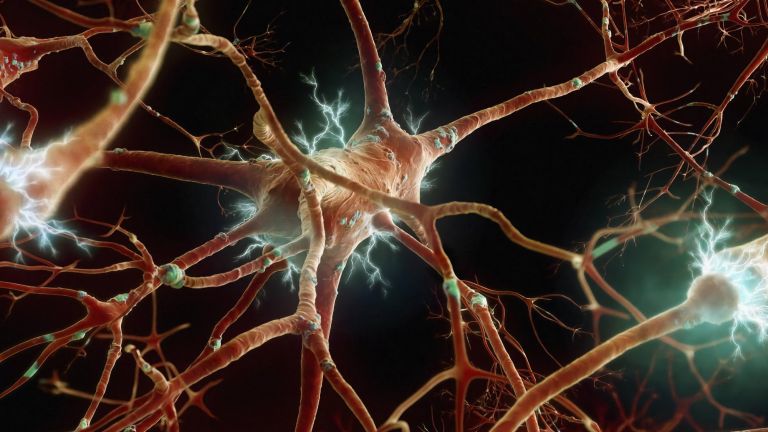
The Epithalamus

The epithalamus, together with the pineal gland, has long captured the imagination of philosophers. Even today, this part of the diencephalon remains a mystery to neuroscientists. It consists mainly of two reins and a cone.
Wissenschaftliche Betreuung: Prof. Dr. Jochen F. Staiger
Veröffentlicht: 09.10.2025
Niveau: mittel
Der Epithalamus fasst in der klassischen Anatomie zwei Strukturen zusammen – zum einen die Epiphyse, die Zirbeldrüse, die über ihr Hormon Melatonin den Schlaf-Wach-Rhythmus beeinflusst. Und zum zweiten die Habenulae, die bei Vermeidung, Belohnung, Stress, Schmerz und sogar der Entscheidungsfindung und Sucht eine wichtige Rolle spielen. Beide Strukturen haben beim Säuger allerdings nichts miteinander zu tun, die alten Anatomen hatten schlicht noch nicht die heutigen Möglichkeiten zu dieser Erkenntnis.
Das Hormon Melatonin aus der Zirbeldrüse steuert den Schlaf-Wach-Rhythmus: Es dockt an Rezeptoren des Nucleus suprachiasmaticus im Hypothalamus – quasi der inneren Uhr des Menschen –, und kurbelt den Schlaf an. Die Zirbeldrüse produziert Melatonin nur bei Dunkelheit, also nachts, und wirkt sozusagen als Zeitgeber im Körper. Es heißt, ältere Menschen schütteten weniger Melatonin aus und benötigten daher weniger Schlaf. Das Hormon spielt auch eine Rolle bei der Entstehung von Jetlag bei Fernreisen und bei körperlichen Problemen durch Schichtarbeit.
Außerdem beeinflusst Melatonin die Keimdrüsen, indem es verhindert, dass die Hypophyse gonadotrope, also keimdrüsenstimulierende, Hormone ausschüttet, darunter das Follikelstimulierende Hormon und das Luteinisierende Hormon. Fällt die Zirbeldrüse bei Kindern aus, kann das eine frühzeitige Pubertät zur Folge haben. Nur am Rand: Bei Amphibien führt Melatonin zu einer Depigmentierung der Haut.
Melatoninhaltige Medikamente sollen gegen den Jetlag wirken, den Schlaf fördern oder freie Radikale abfangen und so Krebs vorbeugen. Viele dieser Wirkungen sind jedoch nicht bewiesen. Die europäische Behörde für Lebensmittelsicherheit (EFSA) befand im Jahr 2010, dass die Behauptung „Melatonin trägt zur Linderung des subjektiven Jetlag-Gefühls bei“ wissenschaftlich gerechtfertigt sei, es aber nicht genug Belege dafür gebe, dass die Einnahme melatoninhaltiger Mittel die Schlafqualität verbessere oder die Einschlafzeit verkürze.
„ ... die Nuclei habenulares ... bilden vermutlich eine Schaltstation zwischen Riechhirn und Hirnstamm“ – so steht es in älteren Lehrbüchern. Doch womöglich müssen diese umgeschrieben werden, denn jüngere Erkenntnisse lassen vermuten, dass die Zügelkerne gar keine olfaktorischen Informationen bekommen.
Dafür tun sich andere – und weitreichendere – Aufgaben für die Habenula auf: Aus den Basalganglien gelangen Informationen über Fehler und Bestrafung zur Habenula, die dann mit dem ventralen Tegmentum und der Substantia nigra Strukturen des dopaminergen Systems beeinflusst – und in dieser Funktion ebenfalls die Motorik hemmt. Zum selben Ergebnis führen auch Informationen über Schmerz und Stress aus dem limbischen System, wobei von Habenula dann zusätzlich zu den dopaminergen Kernen auch die Raphe-Kerne und damit die Serotoninproduktion beeinflusst werden.
Basalganglien
Basalganglien/Nuclei basales/basal ganglia
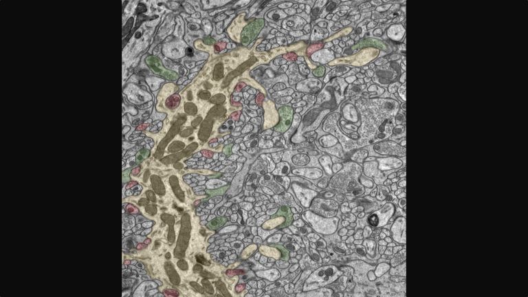
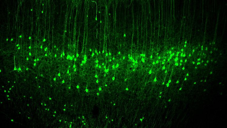
Basalganglien sind eine Gruppe subcorticaler Kerne (unterhalb der Großhirnrinde gelegen) im Telencephalon. Zu den Basalganglien zählen der Globus pallidus und das Striatum, und je nach Autor weitere Strukturen, wie z. B. die Substantia nigra und der Nucleus subthalamicus. Die Basalganglien werden primär mit der Willkürmotorik in Verbindung gebracht, beeinflussen aber auch Motivation, Lernen und Emotion.
Almost hidden on the rear wall of the third ventricle is the epithalamus. It sits behind and above the much larger thalamus, which explains its name: The Greek prefix “epi” means “on.” The epithalamus includes the impressive habenulae – in English, the “reins” – two strands of brain tissue that unite dynamically in the middle, at the unpaired epiphysis, the pineal gland. Viewed from behind, the structure actually resembles the reins of a horse's harness, which are attached to both sides of a bridle.
Three other structures are also considered part of the epithalamus. First, the habenulae continue into the striae medullares, white medullary stripes that run across the thalamus and connect it to the habenulae. Furthermore, the posterior commissure, also known as the epithalamic commissure, and the pretectal area are also considered part of the epithalamus. Commissural fibers always cross from one hemisphere of the brain to the other, and in the case of the posterior commissure, fibers from the tectum and the tegmentum in the midbrain, among others, cross sides. In terms of function, however, both the posterior commissure and the pretectal area belong to the visual system: the pretectal area is at work in the pupillary reflex, for example, when the pupils dilate in the dark or constrict in sudden light.
The official star of the epithalamus is probably the epiphysis, the pineal gland. At least by name, it was already known before the birth of Christ; at that time and later, all kinds of theories surrounded this organ, which is less than a centimeter in size: for example, it was assumed that the pineal gland was a kind of valve for thoughts and memories. The gland is located above the tectum, protruding from the third ventricle and shaped like a pine cone. This is where its Latin name comes from: Glandula pinealis, pineal gland. The epiphysis is largely covered by the inner meninges, which supply blood vessels to the gland. It is mainly composed of pinealocytes, the hormone-producing cells of the glandular tissue. Connective tissue segments the tissue into many vesicles, which look like honeycombs under the microscope when viewed in cross-section. The pineal gland also contains glial cells as support cells and nerve fibers.
Function of the epiphysis
“There is a small gland in the brain in which the soul performs its function more specifically than in any other part of the body,” wrote the philosopher René Descartes about the pineal gland in the 17th century. He believed that body and soul unite in this organ – an idea that is completely divorced from reality. Today we know that the pineal gland produces the hormone melatonin (see box) and releases it into the blood. However, it only does this at night. Daylight inhibits the enzymes that produce melatonin from serotonin in two steps.
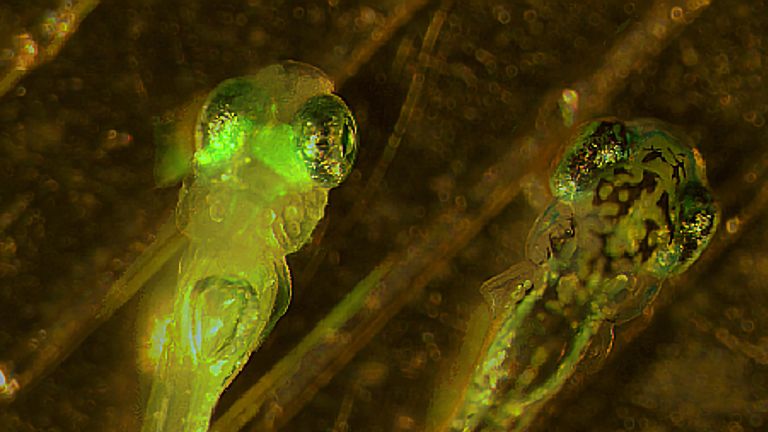
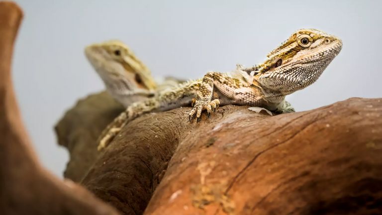
Originally, the pineal gland was not only an endocrine organ, but also a sensory organ with photoreceptor cells. However, these have regressed in the course of evolution. In fact, some amphibians and reptiles, such as the New Zealand tuatara (often referred to as a living fossil), have a third eye under the skin of their skull, the parietal eye. This allows light to fall directly into the brain, enabling the animals to perceive light-dark differences particularly well. In mammals, however, the skull is so thick that the pineal gland no longer needs its photoreceptor cells. Nevertheless, it receives light signals via extensive nerve pathways that run from the retina to the hypothalamus and into the spinal cord, reaching the epiphysis via the superior cervical ganglion, the cervical sympathetic nerve. These are also the only nerve fiber connections to the organ in mammals.
The pineal gland participates in the regulation of the day and night rhythm via melatonin and, in addition to a variety of other internal organs, also influences the gonads. In addition to melatonin, the epiphysis also releases other compounds, neuropeptides, into the blood, the effects of which are still unknown. Even the function of melatonin is still being researched. The pineal gland therefore remains an exciting field of research for neuroscientists today.
Even before the age of 20, i.e. at a relatively young age, the epiphysis begins to calcify. Support cells multiply rapidly, actual glandular tissue dies off, and cysts form in which calcium and magnesium salts are deposited. Doctors call this phenomenon “brain sand” or “acervulus” – these calcium deposits are clearly visible on X-rays. However, their significance is still unclear.


The reins – not just anatomically
Although its shape might suggest otherwise, the habenula is not a strand of fibers, but rather a collection of core areas. The striae medullares, on the other hand, bring fibers from the septal nuclei of the limbic system, the preoptic nuclei, and the amygdala complex into this region. These fibers run into the reins. At the transition between the two lie the nuclei habenulares, the reins nuclei. Despite their small size, they have surprisingly far-reaching functions that can influence the entirety of our behavior, right down to our decision-making.
The lateral habenula influences the reward system by inhibiting it when the result does not meet expectations. It thus also plays a role in corresponding learning processes, the stress response with flight or fight, and even the processing of pain. It even becomes active in fear conditioning. All of this is extremely useful when it comes to adapting to changes in the environment. However, it can also overshoot the mark – if the reward center is inhibited too much, meaning that too little dopamine and serotonin are released, the result can be anhedonia and even depression.
The medial habenula has similar, almost even more far-reaching competencies: it regulates moods, processes negative emotions, and motivates reactions to unpleasant and painful situations. In doing so, it significantly influences higher brain functions such as decision-making and attention. However, it also plays a role in withdrawal and relapse into addiction.
Further reading:
- EFSA's position on melatonin-containing medicines; URL: http://www.efsa.europa.eu/en/efsa-journal/doc/1467.pdf; to the website.
First published on August 28, 2011
Updated on October 9, 2025