More Than Just a Pump

The heart is the hardest-working muscle in the body. To ensure it beats appropriately around the clock throughout a person’s life, it has borrowed a few tricks from the brain and transformed specialized muscle cells into an electrically excitable communication network.
Scientific support: Dr. Ramanan Ganeshan
Published: 01.10.2022
Difficulty: easy
- The heart works at full speed throughout a person’s life. Two weeks after fertilization, it begins to form, and just one week later, it’s already beating.
- Over the course of an 80-year human life, the heart beats nearly three and a half billion times, pumping over 210 million liters of blood.
- The heart serves two circulatory systems simultaneously: systemic circulation and pulmonary circulation. In this way, it sends oxygen-rich blood from the lungs to the body and transports oxygen-poor blood back to the lungs for reoxygenation.
- Muscle cells in the heart do more than just pump blood. Specialized muscle cells act as pacemakers; they generate the electrical impulses that trigger each heartbeat and transmit them.
- The heart regulates its rhythm almost entirely on its own. However, impulses from the Autonomic nervous system adjust the heart rate to meet current needs.
- Cells in the heart and brain are similar in their energy requirements, longevity, and poor ability to regenerate.
- There are numerous interactions between the heart and the brain, and they influence one another.
Autonomic nervous system
The part of the nervous system that primarily controls unconscious vital functions such as breathing, heartbeat, and blood pressure. The autonomic nervous system is divided into the sympathetic nervous system, which is active in performance and stress situations, and the parasympathetic nervous system, which is active during rest and recovery phases. In some cases, the enteric nervous system, which is responsible for gastrointestinal functions, is also considered part of the autonomic nervous system.
The achievements of civilization may be many and varied, but there are well-founded doubts as to whether they are good for the heart. Numerous lifestyle factors in modern industrial societies contribute to conditions such as high blood pressure, obesity, diabetes, dementia, and, of course, cardiovascular disease. Furthermore, they hinder or directly affect the heart’s function.
For hundreds of thousands of years, humans lived in close harmony with the rhythms of the days and seasons, were regularly active, and consumed mostly moderate amounts of minimally processed food. Under these conditions, the heart was likely able to function largely unimpeded and reach its full potential.
Today, various supposed conveniences of modern life place a strain on the cardiovascular system. A lack of physical activity prevents the heart from reaching its full potential. Eating too much food containing excessive amounts of sugar, unhealthy fats, and other harmful components leads to obesity. In an overweight body, this contributes – both directly and indirectly through numerous signaling cascades – to increased inflammation, which can form deposits in blood vessels and have other harmful effects. The disconnection of daily life from natural light rhythms – and the often accompanying reduction in sleep quality and quantity – places additional strain on the system.
Many connections between lifestyle, lifestyle-related diseases, and heart function are still unknown or controversial. But it’s considered likely that changing stress patterns play a major role. It’s no longer the occasional threat to life in the face of a predator or enemy that often plays the biggest role. Instead, constant social stress keeps not only the brain but also the heart in a constant state of alert.
Knowing by heart; words that come from the heart; hearts that are in the mouth or sink to the boots – much of what we mull over in our minds seems closely linked to the organ that beats in our chest. ▸ Aristotle even identified the heart as the seat of the soul. ▸ The brain, on the other hand, he regarded as merely a cooling system. Today we know that the Emotions we feel “in our hearts” are actually generated by storms of neural activity in the brain. However, evidence of parallels ▸ The Heart-Brain Connection and links between the heart and the brain has long been mounting ▸ Heart and Brain – A Dynamic Duo, and these are significant in many biological and medical respects: Mental health and heart health are often closely linked ▸ When Heart and Brain Suffer Together.
Emotions
Neuroscientists understand "emotions" to be complex response patterns that include experiential, physiological, and behavioral components. They arise in response to personally relevant or significant events and generate a willingness to act, through which the individual attempts to deal with the situation. Emotions typically occur with subjective experience (feeling), but differ from pure feeling in that they involve conscious or implicit engagement with the environment. Emotions arise in the limbic system, among other places, which is a phylogenetically ancient part of the brain. Psychologist Paul Ekman has defined six cross-cultural basic emotions that are reflected in characteristic facial expressions: joy, anger, fear, surprise, sadness, and disgust.
The life of a heart
The heart plays a central role in life, almost from the very beginning. Only during the first two weeks after fertilization is the human embryo heartless. Shortly after it implants in the uterus, small cavities inside it fuse into a tube that initially resembles a slug. At the beginning of the fourth week of development, the heart tube begins to pump – already detectable by ultrasound at this stage. Over the next five weeks, the tube coils into a loop shape. Subsequently, the various heart chambers form, and walls and valves develop to separate the chambers from one another. Finally, the major blood vessels take shape.
In just nine weeks, the human heart is essentially fully formed and will continue to function for the rest of the person’s life. While it beats leisurely at the very beginning of development – at a rate similar to the mother’s heart, about 80 times per minute – the unborn baby soon picks up the pace considerably. By the seventh week of development, the developing little heart is racing at 185 beats per minute and then gradually slows down again to an average of 80 beats per minute by the time adulthood is reached.
In healthy adults, each beat pumps about 80 milliliters of blood into the body. That’s roughly 5 liters per minute – about the total volume of blood in the body. When we get excited or exert ourselves, the heart can temporarily reach, depending on age, more than 200 beats per minute and pump up to 20 liters per minute. Using the average adult values as a basis, a person reaches over 115,000 heartbeats and 7,200 liters of pumped blood per day. That amounts to roughly 42 million heartbeats and just under 263,000 liters of blood per year of life, and just under three and a half billion beats and over 210 million liters of blood over the course of an 80-year human life.
The center of the blood circulation
South African surgeon Christiaan Barnard, who performed the first heart transplant in 1967, is said to have remarked: “The heart is just a pump.” Experts no longer view the matter quite so simply today. Yet it is precisely the heart’s pumping function that is central to our lives. We depend on the heart, as the engine of our circulatory system, to perform its duties reliably, around the clock.
The basic principle is simple: The heart consists of two halves separated by the septum.
Each half is further divided into an atrium and a ventricle. Blood can flow through the heart’s halves only in one direction. This is ensured by the heart valves, each of which opens in only one direction. The two halves of the heart serve two different circulatory systems. The systemic circulation supplies the body with oxygen. Oxygen-rich blood flows through the pulmonary veins into the atrium and then into the ventricle of the left half of the heart. With its powerful pumping action, the left ventricle then sends the oxygen-rich blood through the aorta to the most remote, fine blood vessels of the systemic circulation, the capillaries, where the blood releases oxygen and nutrients to the body’s various tissues. On the return journey, the now oxygen-depleted blood flows through the body’s veins back to the heart - into the atrium and ventricle of the right side of the heart. From there, it travels through the pulmonary arteries to the lungs, where it is reoxygenated. This smaller circulation is known as the pulmonary circulation.
Septum
area septalis
The septal nuclei are located medially in the basal forebrain, near the anterior tip of the cingulate gyrus. They are connected to the olfactory cortex and linked to other limbic structures via the fornix. Functionally, they play a role in emotional processes and reward processing.
Workhorses and pacesetters
To ensure that the heart can supply every corner of the body during continuous operation – and always precisely tailored to current needs – it possesses a number of special characteristics that, in some cases, show astonishing parallels to the brain. In simple terms, there are essentially two types of cells in the heart: the cells of the working muscle and the cells of the conduction system. As the name suggests, the working muscle ensures that the heart contracts reliably, powerfully, and rhythmically. Unlike skeletal muscle, whose cells fuse into long muscle fibers with multiple cell nuclei even before birth, heart muscle cells retain their individuality. They typically contain no more than two cell nuclei, which are formed during a final round of DNA replication before the cell leaves the cell cycle permanently.
Heart muscle cells are by no means lone fighters, however. Rather, they link up with their neighbors via branched extensions, thereby also forming long muscle fibers. At the connection points are many small pores known as gap junctions. Through these, cytoplasm can flow directly from cell to cell. This allows not only chemical messengers but also, through the exchange of ions, electrical signals to be transmitted particularly quickly. This is useful when, as in the heart muscle, the rhythmic activity of many cells needs to be coordinated. A similar mechanism is found in the brain; here, too, gap junctions connect specific nerve cells that work closely together, such as in the hippocampus, the central hub between short-term and long-term memory.
The cardiac conduction system exhibits even more parallels to the brain. It consists of modified heart muscle cells that interconnect to form a complex signaling network equipped with various backup mechanisms. This network is responsible for generating and transmitting the electrical impulses that set the rhythm for the working muscles – and it does so almost entirely without the involvement of the nervous system. Cardiac muscle cells in the cardiac conduction system are less able to contract than their counterparts in the working muscle, but they are specialists in generating and transmitting electrical impulses, known as action potentials.
The cardiac conduction system consists of several components. The primary pulse generators are the pacemaker cells in the sinus node. This is a spindle-shaped structure about one to two centimeters long located in the wall of the right atrium. The pacemaker cells spontaneously, and ideally at regular intervals, generate the electrical impulses that initiate each heartbeat. This works because pacemaker cells have the ability to repeatedly depolarize themselves, thereby rhythmically generating new action potentials. Each Action potential in electrically excitable cells is followed by a repolarization phase, during which the sodium channels in the cell membrane close, halting the further influx of positively charged sodium ions. At the same time, positively charged potassium ions are pumped out of the cell. As a result, the charge inside the cell gradually becomes more negative again, and the cell approaches its Resting potential Most electrically excitable cells, including neurons and cells of the heart muscle, now remain at their resting potential for a while. The pacemaker cells in the heart’s sinus node are different; they possess very special sodium channels that reopen immediately upon reaching hyperpolarization. The interior of the cell immediately becomes more positive again, leading to renewed depolarization.
Thanks to this special mechanism, the pacemaker cells in the sinus node can function as autonomous pacemakers. However, they are not entirely independent of the rest of the body. In particular, the Autonomic nervous system can use its neurotransmitters to both lower and raise the firing rate of the sinus node – and thus the overall heart rate. The regulation of calcium Homeostasis plays an important role in this process – another similarity with the brain, where calcium-dependent signals play a variety of roles in modulating synapses and neural networks.
From the sinoatrial node, the impulses travel to the atrioventricular node, which is located in the wall between the right and left atria near the border with the ventricles, and on to the so-called His bundle. It runs in two strands (Tawara branches) along the cardiac Septum toward the apex of the heart. There, they branch into the final component of the cardiac conduction system, the so-called Purkinje fibers. These are connected to the cell chains of the cardiac muscle and directly dictate their rhythm. Interestingly, the cells located downstream of the sinoatrial node in the cardiac conduction system can also function as pacemakers. Under normal conditions, however, they do not come into play; they can, for example, take over if the sinoatrial node fails.
Action potential
In excitable cells (e.g., neurons or muscle cells), very rapid changes in electrical potential occur across the cell membrane. This event is the basis for signal conduction along the axon of the nerve cell. The action potential continues along the cell membrane and, according to the all-or-nothing principle, only occurs when the cell has been sufficiently excited.
Resting potential
Resting membrane potential
The membrane potential of a neuron at rest. The inflow and outflow of ions are in equilibrium. It ranges from –50 to –100 mV.
Autonomic nervous system
The part of the nervous system that primarily controls unconscious vital functions such as breathing, heartbeat, and blood pressure. The autonomic nervous system is divided into the sympathetic nervous system, which is active in performance and stress situations, and the parasympathetic nervous system, which is active during rest and recovery phases. In some cases, the enteric nervous system, which is responsible for gastrointestinal functions, is also considered part of the autonomic nervous system.
Homeostasis
Homeostasis is the ability of an organism to maintain internal conditions such as metabolism, body temperature, blood pressure, etc. at a relatively constant level. The maintenance of homeostasis is controlled by the autonomic nervous system, meaning that humans cannot directly influence its regulation at will.
Septum
area septalis
The septal nuclei are located medially in the basal forebrain, near the anterior tip of the cingulate gyrus. They are connected to the olfactory cortex and linked to other limbic structures via the fornix. Functionally, they play a role in emotional processes and reward processing.



Energy demands and regenerative capacity
The constant generation and transmission of electrical impulses, as well as the tireless pumping action of the cardiac muscle, are energetically costly. To ensure an adequate energy supply, cardiac muscle cells therefore contain a particularly large number of Mitochondria. This is another parallel to the brain, whose nerve cells are also very energy hungry. Both organs share yet another commonality: the vast majority of their cells commit to their specialized tasks shortly after birth at the latest – and do so so rigorously that they lose the ability to divide. This means that both tissues are barely capable of regenerating after injury or disease, such as a heart attack.
For a long time, it was thought that the human heart was incapable of regeneration at all. It is now clear that regeneration is indeed possible, but not to an extent that would allow it to repair even major damage. Why this is the case, and why other organisms, such as the zebrafish, have no trouble forming new heart tissue, remains unclear to this day.
It is conceivable that this inability is directly related to the high energy demands of the heart and brain. Where particularly large numbers of mitochondria convert energy-rich substances into ATP, the body’s universal energy currency, with the help of oxygen, more free radicals are also produced. Due to their high chemical reactivity, these free radicals can damage DNA. In cells that are still dividing, this implies an increased risk of tumors. The energy-intensive functioning of the heart and brain may therefore not be compatible with rapid cell division.
Mitochondria
Mitochondria are organelles inside a cell. They are also referred to as the "powerhouse" of cells because they supply them with energy. They have their own DNA, which is only inherited from the mother.
Interactions
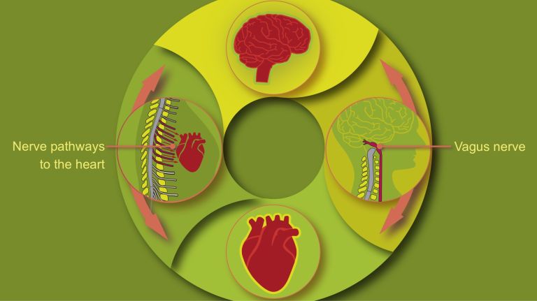
The similarities between the heart and the brain become all the more interesting the closer one looks at the diverse connections and interactions between the two organs. It is now well established that mental illnesses and heart disease are closely linked. If the heart is not functioning properly, problems with the brain’s blood supply can quickly lead to impairments in its functions, such as Dementia. Conversely, not only can intense physical activity push the heart to its limits, but severe emotional stress can also threaten its functions ▸ How Mental Stress in the Mind Harms the Heart. Exactly how the interaction between the two systems works, and which factors can throw them out of balance, is therefore increasingly the subject of research. It is becoming increasingly clear that the heart is ultimately neither the seat of the soul nor a mere pump, but rather a bit of both.
Dementia
Dementia
Dementia is an acquired deficit of cognitive, social, motor, and emotional abilities. The most well-known form is Alzheimer's disease. "De mentia" means "without mind" in English.
Further reading
- Koehler U, Hildebrandt O, Hildebrandt W, Aumüller G. The Heart and Cardiovascular System from a (Cultural) Historical Perspective [Historical (cultural) view of the heart and cardiovascular system]. Herz. 2021;46(Suppl 1):33-40. doi:10.1007/s00059-020-04914-2 (full text)
- Elhelaly, Waleed M et al.: Redox Regulation of Heart Regeneration: An Evolutionary Tradeoff. Frontiers in Cell and Developmental Biology vol. 4, 137. Dec. 15, 2016, doi:10.3389/fcell.2016.00137 (Full text)
- Islam MR, Lbik D, Sakib MS, et al. Epigenetic Gene expression links heart failure to Memory impairment. EMBO Mol Med. 2021;13(3):e11900. doi:10.15252/emmm.201911900 (Full text)
Gene
Information unit on DNA. Specialized enzymes translate the core component of a gene into ribonucleic acid (RNA). While some ribonucleic acids perform important functions in the cell themselves, others specify the order in which the cell should assemble individual amino acids into a specific protein. The gene thus provides the code for this protein. In addition, a gene also includes regulatory elements on the DNA that ensure that the gene is read exactly when the cell or organism actually needs its product.
Memory
Memory is a generic term for all types of information storage in the organism. In addition to pure retention, this also includes the absorption of information, its organization, and retrieval.