The Cerebellar Peduncles

The cerebellum is neither an appendage nor isolated organ, but is closely connected to the rest of the nervous system: three thick bundles of fibers anchor it to the brain stem. They act like powerful cables through which a constant stream of information flows into the cerebellum – and, to a lesser extent, out again.
Scientific support: Prof. Dr. Hans-Dieter Hofmann, Prof. Dr. Andreas Vlachos
Published: 20.09.2025
Difficulty: intermediate
The Cerebellum is connected to the rest of the central nervous system via three thick bundles of fibers. The upper and lower Cerebellar peduncles carry information from the body to the cerebellum and transmit signals for movement correction to the Brain stem and cerebral Cortex. The middle cerebellar peduncle contains only afferent fibers that transmit signals from the cerebral cortex.
Cerebellum
Cerebellum
The cerebellum is an important part of the brain, located at the back of the brain stem and below the occipital lobe. It consists of two cerebellar hemispheres covered by the cerebellar cortex and plays an important role in motor processes, among other things. It develops from the rhombencephalon.
Cerebellar peduncles
pedunculi cerebelli
Three fiber connections on the right and left sides that connect the cerebellum to the brain stem. All afferent and efferent fibers of the cerebellum run through these connections.
Brain stem
truncus cerebri
The "trunk" of the brain, to which all other brain structures are "attached," so to speak. From bottom to top, it comprises the medulla oblongata, the pons, and the mesencephalon. It transitions into the spinal cord below. It is a center for vital functions such as breathing and heartbeat and contains ascending and descending pathways between the cerebrum, cerebellum, and spinal cord.
Cortex
cortex cerebri
Cortex refers to a collection of neurons, typically in the form of a thin surface. However, it usually refers to the cerebral cortex, the outermost layer of the cerebrum. It is 2.5 mm to 5 mm thick and rich in nerve cells. The cerebral cortex is heavily folded, comparable to a handkerchief in a cup. This creates numerous convolutions (gyri), fissures (fissurae), and sulci. Unfolded, the surface area of the cortex is approximately 1,800cm².
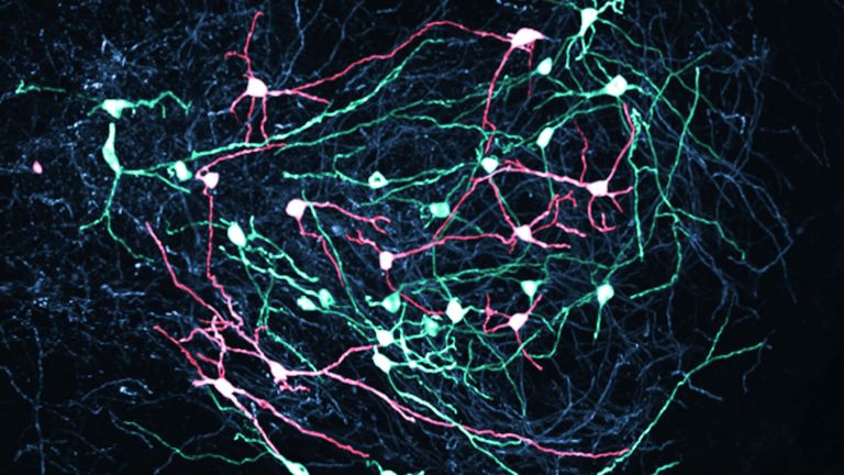
If the Cerebellum is the “tree of life” due to its branched interior, then the Cerebellar peduncles are the roots that anchor the cerebellum firmly to the pons, extended spinal cord, and neighboring brain regions. The three thick bundles of fibers are direct extensions of the White matter of the cerebellum. They transport information from all possible parts of the body and carry outgoing signals to other brain regions. Like powerful cables, the cerebellar peduncles connect the otherwise isolated cerebellum to the rest of the brain.
The cerebellum can be thought of as a major consumer of information, as there are significantly more fibers that bring information in than those that carry signals out of the cerebellum: there is one outgoing fiber for every 40 incoming fibers. After all, the cerebellum needs to keep track of what is happening in the body at all times. To do this, it needs a great deal of input, for example from the vestibular system, receptors of proprioception, spinal cord, and motor cortex, both about movements that are currently taking place and those that are being planned in the cerebral Cortex. Once it has coordinated the various signals, the corrective output that is the result of its workrequires significantly less transmission capacity. In short, the cerebellum thrives on input – only in this way can it continuously monitor movements and adjust them with millimeter precision.
Cerebellum
Cerebellum
The cerebellum is an important part of the brain, located at the back of the brain stem and below the occipital lobe. It consists of two cerebellar hemispheres covered by the cerebellar cortex and plays an important role in motor processes, among other things. It develops from the rhombencephalon.
Cerebellar peduncles
pedunculi cerebelli
Three fiber connections on the right and left sides that connect the cerebellum to the brain stem. All afferent and efferent fibers of the cerebellum run through these connections.
White matter
The white matter refers to the myelinated fibers of the nervous system that connect one neuron to another. The white color is caused by the myelin sheath surrounding the fibers.
Cortex
cortex cerebri
Cortex refers to a collection of neurons, typically in the form of a thin surface. However, it usually refers to the cerebral cortex, the outermost layer of the cerebrum. It is 2.5 mm to 5 mm thick and rich in nerve cells. The cerebral cortex is heavily folded, comparable to a handkerchief in a cup. This creates numerous convolutions (gyri), fissures (fissurae), and sulci. Unfolded, the surface area of the cortex is approximately 1,800cm².
Location and structure
Whoever once gave the three Cerebellar peduncles their names was not very imaginative: they are simply called the upper, middle, and lower peduncles, or in Latin, pedunculus cerebellaris superior, medius, and inferior.
The upper stem connects the Cerebellum to the midbrain, the middle stem to the pons, and the lower stem to the extended spinal cord, the Medulla oblongata A thin layer of white matter, the velum medullare superius, stretches between the upper cerebellar peduncles. It forms the roof of the fourth ventricle and borders the Tectum of mesencephalon at the rear.
The middle cerebellar peduncle is located further to the side. It is the strongest of the three fiber bundles, and, unlike the other two peduncles, contains only afferent fibers. It developed later in evolution and has pushed itself between the upper and lower cerebellar peduncles. In the process, it also pushed apart some fiber bundles that belong together functionally, namely the pathways that carry information from the Spinal cord As a result, only some of the fibers from the spinal cord to the cerebellum now run through the inferior cerebellar peduncle, while the rest of this pathway is routed through the superior cerebellar peduncle. Medical students are explicitly taught this as an exception to the rule in exam swoting books. Otherwise, the pathways can be logically assigned to the three cerebellar peduncles according to their place of origin.
Cerebellar peduncles
pedunculi cerebelli
Three fiber connections on the right and left sides that connect the cerebellum to the brain stem. All afferent and efferent fibers of the cerebellum run through these connections.
inferior
An anatomical position designation – inferior means located further down, the lower part.
Cerebellum
Cerebellum
The cerebellum is an important part of the brain, located at the back of the brain stem and below the occipital lobe. It consists of two cerebellar hemispheres covered by the cerebellar cortex and plays an important role in motor processes, among other things. It develops from the rhombencephalon.
Medulla oblongata
Area of the brain that transitions into the spinal cord. The medulla oblongata comprises nerve pathways between the spinal cord and higher brain regions, as well as numerous core areas with functions that are in some cases vital, such as breathing, heartbeat, and certain reflexes.
Tectum
A structure in the midbrain consisting of two pairs of mounds, the upper colliculi and the lower colliculi.
Spinal cord
medulla spinalis
The spinal cord is the part of the central nervous system located in the spine. It contains both the white matter of the nerve fibers and the gray matter of the cell nuclei. Simple reflexes such as the knee-jerk reflex are already processed here, as sensory and motor neurons are directly connected. The spinal cord is divided into the cervical, thoracic, lumbar, and sacral spinal cord.


Incoming fibers: What goes in?
Most signals reach the Cerebellum via the middle cerebellar peduncle. It is the largest bundle of fibers and contains only incoming pathways. These ultimately originate in the cerebral cortex, are connected in the pons, and provide the cerebellum with information about the movements the Cerebrum is currently planning.
Most of the information from the rest of the body otherwise runs through the inferior cerebellar peduncle: it informs the cerebellum, for example, about the position of a leg, the trunk, or an arm, how they are moving, and what the Vestibular system is reporting. Specifically, it carries fibers from the vestibular nuclei, the brainstem, the spinal cord, and the inferior olivary body.
Finally, there is the upper cerebellar peduncle. It not only transmits parts of the Spinal cord pathways, but also signals from nuclei in the midbrain, such as the Nucleus ruber. It thus supplements the information from the spinal cord and brain stem, and provides the cerebellum with an even more complete picture of the body's posture and movement.
All fibers that reach the cerebellum end as climbing fibers or mossy fibers in the cerebellar cortex, where they are integrated into the dense network of circuits and further processed.
Cerebellum
Cerebellum
The cerebellum is an important part of the brain, located at the back of the brain stem and below the occipital lobe. It consists of two cerebellar hemispheres covered by the cerebellar cortex and plays an important role in motor processes, among other things. It develops from the rhombencephalon.
Cerebrum
telencephalon
The cerebrum comprises the cerebral cortex (gray matter), the nerve fibers (white matter), and the basal ganglia. It is the largest part of the brain. The cortex can be divided into four cortical areas: the temporal lobe, frontal lobe, occipital lobe, and parietal lobe.
Its functions include the coordination of perception, motivation, learning, and thinking.
inferior
An anatomical position designation – inferior means located further down, the lower part.
Vestibular system
Vestibular apparatus/Organon vestibulare/vestibular organ
The vestibular system is part of the inner ear. Its sensors are located in the semicircular canals. As part of the balance system, it detects circular movements (rotations), acceleration, and gravity.
Spinal cord
medulla spinalis
The spinal cord is the part of the central nervous system located in the spine. It contains both the white matter of the nerve fibers and the gray matter of the cell nuclei. Simple reflexes such as the knee-jerk reflex are already processed here, as sensory and motor neurons are directly connected. The spinal cord is divided into the cervical, thoracic, lumbar, and sacral spinal cord.
Nucleus
In cell biology, the nucleus in a cell is the cell nucleus, which contains the chromosomes, among other things. In neuroanatomy, the nucleus in the nervous system refers to a collection of cell bodies – known as gray matter in the central nervous system and ganglia in the peripheral nervous system.
Output fibers: What goes out?
The signals processed by the Cerebellum travel from its Cortex to the Cerebellar nuclei inside. Almost all fibers that leave the cerebellum originate here; only a few originate directly in the cerebellar cortex.
Most of the output fibers leave the cerebellum via the upper peduncle. These pathways lead to motor centers in the brain stem, to the red nucleus, and via the thalamus also to the cerebral cortex. A smaller portion of the outgoing fibers runs back to the Brain stem via the inferior peduncle, including to the vestibular nuclei and the inferior olivary body. In this way, the cerebellum can also regulate its own inputs and at the same time influence the balance system.
A special feature is the mode of action of these pathways: they run ipsilaterally, i.e., they act on the same side of the body – the right Hemisphere of the cerebellum controls the right side of the body, and the left hemisphere controls the left side. This distinguishes the cerebellum from the cerebrum, in which each hemisphere primarily controls the opposite side of the body, i.e., contralaterally.
Cerebellum
Cerebellum
The cerebellum is an important part of the brain, located at the back of the brain stem and below the occipital lobe. It consists of two cerebellar hemispheres covered by the cerebellar cortex and plays an important role in motor processes, among other things. It develops from the rhombencephalon.
Cortex
cortex cerebri
Cortex refers to a collection of neurons, typically in the form of a thin surface. However, it usually refers to the cerebral cortex, the outermost layer of the cerebrum. It is 2.5 mm to 5 mm thick and rich in nerve cells. The cerebral cortex is heavily folded, comparable to a handkerchief in a cup. This creates numerous convolutions (gyri), fissures (fissurae), and sulci. Unfolded, the surface area of the cortex is approximately 1,800cm².
Cerebellar nuclei
A group of four paired nuclei located in the white matter of the cerebellum: the dentate nucleus, emboliform nucleus, globose nucleus, and fastigial nucleus. Functionally, the cerebellar nuclei are associated with motor tasks.
Brain stem
truncus cerebri
The "trunk" of the brain, to which all other brain structures are "attached," so to speak. From bottom to top, it comprises the medulla oblongata, the pons, and the mesencephalon. It transitions into the spinal cord below. It is a center for vital functions such as breathing and heartbeat and contains ascending and descending pathways between the cerebrum, cerebellum, and spinal cord.
inferior
An anatomical position designation – inferior means located further down, the lower part.
Hemisphere
The cerebrum and cerebellum each consist of two halves – the right and left hemispheres. In the cerebrum, they are connected by three pathways (commissures). The largest commissure is the corpus callosum.
First published on August 23, 2011
Last updated on September 20, 2025