Perfect Timing
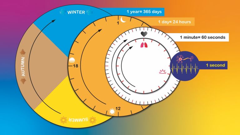
From milliseconds to seasons – the nervous system and the body’s organs determine the rhythms of our lives. To do this, the organism has a highly complex system of internal clocks, pacemakers, and correction mechanisms.
Scientific support: Prof. Dr. Henrik Oster
Published: 02.05.2024
Difficulty: easy
- Rhythms dominate our bodies. They follow the cycle of day and night but can be influenced by many factors.
- The supreme authority of the internal clock is the Suprachiasmatic nucleus (SCN), a pair of brain nuclei containing a total of about 50,000 nerve cells located above the optic chiasm.
- Most cells in the body follow a 24-hour rhythm, which is driven independently of the SCN by a clock Gene and its associated gene product “per” and is based on the principle of negative feedback.
- The key to identifying processes that occur simultaneously lies in synaptic integration. This allows individual neurons to recognize whether signals from their neighbors belong together.
Suprachiasmatic nucleus
nucleus suprachiasmaticus
A nucleus of the hypothalamus that plays a central role in circadian rhythms, including the sleep-wake cycle. It is the master clock, the body's most important internal clock, controlling melatonin production in the epiphysis. It receives direct input from the retinal ganglion cells.
Gene
Information unit on DNA. Specialized enzymes translate the core component of a gene into ribonucleic acid (RNA). While some ribonucleic acids perform important functions in the cell themselves, others specify the order in which the cell should assemble individual amino acids into a specific protein. The gene thus provides the code for this protein. In addition, a gene also includes regulatory elements on the DNA that ensure that the gene is read exactly when the cell or organism actually needs its product.
“Take this pill three times a day after meals.” Such medical instructions sound familiar, yet given the multitude of bodily functions that follow very different rhythms throughout the day, they also seem rather general. Shouldn’t it be possible to achieve better results through more targeted timing of medication administration?
The question is not new and was raised as early as the 1990s Francis Levi, now emeritus director of cancer research at Paris-Saclay University. At that time, it was already known that, for example, the chemotherapy drug Adriamycin is best tolerated in the early morning, whereas Cisplatin is best tolerated in the late afternoon. In clinical practice, however, it did not seem feasible to administer an infusion to the same patient at 4 a.m., the next at noon, and the third at 10 p.m. Instead, a 24-hour infusion was used, delivering all medications simultaneously.
Levi’s proposed improvement, which he presented in 1994 at the annual meeting of the American Society of Clinical Oncology, consisted of a smart pump that patients carried with them. It automatically administered precise doses of the chemotherapy drugs and, in a study involving 186 patients with advanced colorectal cancer, caused significantly fewer side effects than the standard method. As a result, a greater total dose of the active ingredient could be administered, and the tumors of those treated in this way shrank more significantly and twice as often. Disappointment followed three years later when Levi published the complete data: For the most important outcome – the survival rate – there was no significant
A similar, larger study failed in 2006. Thus, aligning medication with the body’s internal clock remained a nice theory whose benefits could only be demonstrated in a few cases under practical conditions – such as in some patients with extreme blood pressure fluctuations. But that is not the end of the story.
Anyone who wants to be successful in life should know their priorities and be in the right place at the right time. Today, such wisdom is often taught in management training courses, yet humans have carried the prerequisites for it within us for millions of years: It is our internal clock that gets us out of bed in the morning, guides us through every day from cradle to grave, and ensures the necessary rest periods at night.
Not only do our sleep-wake cycle and seasonal adaptations depend on this internal clock, but also learning and memory, as well as nearly all physiological daily rhythms such as fluctuations in blood pressure, heart rate, Hormone levels, respiratory rate, and even blood clotting.
In fact, there is no single internal clock. Instead, a highly complex, hierarchical system of interconnected timers, pacemakers, and correction mechanisms governs our time. It consists of specific regions of the nervous system, specialized cells, hormones, neurotransmitters, proteins, and nucleic acids. Ultimately, every single cell has its very own internal clock. “We have a veritable clock shop,” explains Gregor Eichele, who heads the “Genes and Behavior” department at the Max Planck Institute for Biophysical Chemistry in Göttingen.
Hormone
Hormones are chemical messengers in the body. They serve to transmit information between organs and cells, usually slowly, e.g., to regulate blood sugar levels. Many hormones are produced in glandular cells and released into the blood. At their destination, e.g., an organ, they dock at binding sites and trigger processes inside the cell. Hormones have a broader effect than neurotransmitters; they can influence various functions in many cells of the body.
Linked to the alternation of day and night
The nervous system’s primary timekeeping mechanism is surprisingly small: the Suprachiasmatic nucleus (SCN), located on both sides of the brain, is part of the Hypothalamus and sits above the crossing of the optic nerves. It contains only about 50,000 neurons – less than three millionths of the total number of our 65 billion nerve cells. Among these are the circadian pacemaker neurons. A defining feature is the strong fluctuations between day and night in the frequency of their spontaneous discharges. Both the excitatory and inhibitory signals for a robust daily rhythm arise from the temporally coordinated activity with which sodium and potassium ions flow through the membranes of the pacemaker neurons, and from the specific change in ionic conductance during action potentials. The pacemaker neurons in the SCN are not only necessary but also sufficient to drive the circadian rhythm. Furthermore, they counteract deviations from the 24-hour day-night rhythm and synchronize our activities with the rotating earth.
In fact, the human circadian clock would actually have a rhythm of just under 25 hours, as is known from experiments with volunteers who lived in uniformly lit rooms for several weeks. It is thanks to the SCN’s synchronization with real daylight that these subjects quickly regained their rhythm after the experiment ended. Thus, even in the age of artificial lighting, the SCN prevents us from losing our sense of time and turning into zombies after a night of partying.
But how does information about the length of the day reach the SCN? Unsurprisingly, the Eye plays a crucial role in this process. Located in the Retina between the Cones and Rods are specialized Ganglion cells (RGCs), which, alongside the cones and rods, form a third class of Retinal Photoreceptors. They are equipped with the Photopigment melanopsin, which allows them to detect incoming light. The fibers of the RGCs then run to the SCN and other brain regions – including those that regulate mood. Even in people who have gone blind due to damage to the visual cortex, this adjustment of the internal clock functions via the pathway from the RGCs to the SCN – as long as the retina and the nerve pathways extending from it remain intact.
However, nature did not anticipate the invention of artificial light by humans, and in particular of cell phones, tablets, and similar devices. These emit – similar to normal daylight – abundant blue light, to which melanopsin reacts sensitively. The problem: In the evening, the internal clock is particularly sensitive to light effects. If electronic devices are not turned off in the evening, there is a risk of the internal clock being delayed, and the risk of sleep disorders increases.
Suprachiasmatic nucleus
nucleus suprachiasmaticus
A nucleus of the hypothalamus that plays a central role in circadian rhythms, including the sleep-wake cycle. It is the master clock, the body's most important internal clock, controlling melatonin production in the epiphysis. It receives direct input from the retinal ganglion cells.
Hypothalamus
The hypothalamus is considered the center of the autonomic nervous system, meaning it controls many motivational states and regulates vegetative aspects such as hunger, thirst, and sexual behavior. As an endocrine gland (which, unlike an exocrine gland, releases its hormones directly into the blood without a duct), it produces numerous hormones, some of which inhibit or stimulate the pituitary gland to release hormones into the blood.In this function, it also plays an important role in the response to pain and is involved in pain modulation.
excitatory
Exciting synapses are described as excitatory when they depolarize the subsequent cell membrane and can thus lead to the formation of an action potential. An excitatory effect is usually produced by an exciting transmitter (messenger substance), such as glutamate. The opposite is an inhibitory synapse.
Eye
bulbus oculi
The eye is the sensory organ responsible for perceiving light stimuli – electromagnetic radiation within a specific frequency range. The light visible to humans lies in the range between 380 and 780 nanometers.
Retina
The retina is the inner layer of the eye covered with pigment epithelium. The retina is characterized by an inverse (reversed) arrangement: light must first pass through several layers before it hits the photoreceptors (cones and rods). The signals from the photoreceptors are transmitted via the optic nerve to the processing areas of the brain. The reason for the inverse arrangement is the evolutionary development of the retina, which is a protrusion of the brain.
The retina is approximately 0.2 to 0.5 mm thick.
Cones
The cones are a type of photoreceptor in the retina. The three different types of cones – S, M, and L – are each stimulated by short, medium, and long wavelengths of visible light, enabling color vision. They are highly concentrated in the fovea and enable sharp vision.
Rods
The rods are light-sensitive cells with high light sensitivity. They react even to weak light and are therefore responsible for scotopic vision, black-and-white vision, and vision at dusk. The rods are concentrated in the outer areas of the retina and therefore do not provide high visual acuity.
Ganglion
Term for a cluster of nerve cell bodies in the peripheral nervous system. The term nerve node is often used because of its appearance. (Greek gágglion = knot-like)
Retinal
A chemical synthesized from vitamin A. Together with opsin, it forms rhodopsin.
Photoreceptors
Photoreceptors are the light-sensitive cells of the retina; they convert light into electrical potentials. There are approximately 127 million photoreceptors in the retina, including seven million cones and 120 million rods.
Photopigment
Photopigments are light-sensitive molecules in the receptors of the retina. When photons (particles of light) strike the photopigment, it isomerizes, triggering a cascade of various processes. In this way, light is converted into a nerve impulse. All rods have the same type of photopigment, while the cones have one of three possible photopigments.
A clock gene regulates itself
hronobiologists have learned a great deal from dissecting these circuits. However, it is primarily molecular biologists who have discovered what makes the clock tick. They found that an approximately 24-hour cycle also runs inside most of the cells in our bodies. Although this has been studied primarily in fruit flies and mice, and the details as well as the naming of the components differ in part between the various experimental organisms, the structure of this clockwork is apparently very similar throughout the animal kingdom and is even found in the single-celled baker’s yeast.
This periodicity is achieved through the principle of negative feedback: A “clock gene” is read by the cell’s machinery, and messenger RNA (mRNA) is produced from this information, which in turn is translated into a specific protein. During the day, this CLOCK protein activates another clock Gene called “per” (for period). The corresponding per-mRNA is thus used to synthesize the PER protein. Together with other tissue-specific proteins, the PER protein forms a complex that binds to the CLOCK protein and thereby blocks it. The result is that no new per-mRNA is produced, the remaining PER protein gradually breaks down, and the blockage of the CLOCK protein is thus lifted. The cycle then begins anew.
In detail, this system is far more complex: with its fluctuating concentrations, different binding partners, and through equilibrium reactions with other cellular components, PER can, for example, indirectly sense the cell’s energy supply, influence metabolism, or modulate stress responses.
Although the SCN acts as the highest control authority regulating the timing of bodily functions, most organs and tissues can maintain their own rhythm independently thanks to the clock/per system, as can be observed in isolated liver, lung, or kidney cells in culture dishes.
Gene
Information unit on DNA. Specialized enzymes translate the core component of a gene into ribonucleic acid (RNA). While some ribonucleic acids perform important functions in the cell themselves, others specify the order in which the cell should assemble individual amino acids into a specific protein. The gene thus provides the code for this protein. In addition, a gene also includes regulatory elements on the DNA that ensure that the gene is read exactly when the cell or organism actually needs its product.
Rhythms of the body
It makes perfect sense that many internal organs, as well as muscles, adipose tissue, and blood vessels, have a certain degree of independence from the SCN. This allows them to better adapt to changing environmental influences throughout the day thanks to their own molecular clocks. For example, a rising body temperature prepares us for the day even before we wake up, and in the morning, the stress Hormone Cortisol is released in greater quantities. This increases both physical and mental performance.
If necessary, these oscillations are fine-tuned by the SCN to maintain a 24-hour rhythm. There are evidently many cross-connections between the systems, though the details have not yet been fully explored. Only recently was a fast data pathway discovered, thanks to which the so-called mitral cells in the brain’s olfactory center register rhythmic changes in blood vessels caused by the heartbeat. Because more of these “heartbeat sensors” are distributed throughout the entire brain, researchers led by Luna Jammal Salahmeh at the Zoological Institute of the University of Regensburg speculate that they have found an interface here through which the heartbeat could directly influence our thoughts.
Hormone
Hormones are chemical messengers in the body. They serve to transmit information between organs and cells, usually slowly, e.g., to regulate blood sugar levels. Many hormones are produced in glandular cells and released into the blood. At their destination, e.g., an organ, they dock at binding sites and trigger processes inside the cell. Hormones have a broader effect than neurotransmitters; they can influence various functions in many cells of the body.
Cortisol
A hormone produced by the adrenal cortex that is primarily an important stress hormone. It belongs to the group of glucocorticoids and influences carbohydrate and protein metabolism in the body, suppresses the immune system, and acts directly on certain neurons in the central nervous system.

Choreography of brain waves
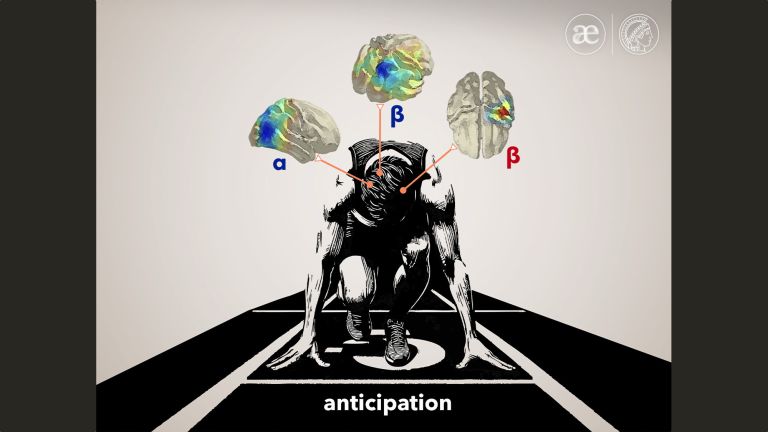
Neurons located beneath the skull in the Neocortex exhibit a completely different rhythm. Cells from different regions can work together, revealing this in the form of “brain waves” that can be visualized using an electroencephalogram (EEG). Since the 1920s, the EEG has therefore become an important research tool, and it is now also used to diagnose neurological and psychiatric disorders.
Generally, the following waveforms are distinguished, sorted by the level of activity:
- Gamma waves with a frequency between 30–100 oscillations per second (hertz) during states of concentration or learning, Beta waves ranging from 12 to 30 hertz, which are generated during normal wakefulness, Alpha waves at 8 to 13 hertz when the subject lies relaxed with their eyes closed,
- theta waves at 4 to 7 hertz, which are generated during dreaming but also indicate memory-related activity, as well as
- delta waves at less than 1 to 4 hertz, which occur during deep sleep.
Between wakefulness and sleep, these brain waves alternate in a fairly clear and consistent pattern. The curves in the EEG, which are still rather “restless” before falling asleep, appear to gradually calm down in the first hour of sleep. The muscles and eyes relax, heart rate, blood pressure, and body temperature drop, and characteristic slow waves with high amplitude can be seen in the EEG – hence the term “slow wave.”
After the sleeper remains at the lowest point of this decline for about half an hour, the EEG waves also gradually become faster again. Similar to when waking up, the eyes begin to move, which has given this sleep phase the name REM sleep (Rapid Eye Movement). The muscles remain paralyzed, but significant variability in heart rate, blood pressure, and body temperature indicates that a dream phase is now taking place. This first dream phase lasts about 10 to 15 minutes, during which men, incidentally, often experience an erection.
These alternating cycles of deep sleep and REM sleep typically repeat about four more times during the rest of the night. As the night progresses, the deep Sleep phases generally become shorter and the REM phases longer until waking. So we already know quite a lot about this phenomenon – though it is still not entirely clear what this phase is actually for!
Neocortex
The neocortex is the phylogenetically youngest part of the cerebral cortex. Since it is structured relatively uniformly in six layers, it is also referred to as the isocortex.
EEG
An electroencephalogram, or EEG for short, is a recording of the brain's electrical activity (brain waves). Brain waves are measured on the surface of the head or using electrodes implanted in the brain itself. The time resolution is in the millisecond range, but the spatial resolution is very poor. The discoverer of electrical brain waves and EEG is the neurologist Hans Berger (1873−1941) from Jena.
Beta waves
Electrical activity of the brain (brain waves) measured on the surface of the head or using electrodes implanted in the brain itself. If the frequency range of the measured activity is between 13 and 30 Hz, these are called beta waves. They are associated with normal waking states and alertness.
Alpha waves
Neuroscientists distinguish between different types of brain waves based on their frequency. Alpha waves oscillate in the mid-frequency range between approximately 8 and 12 hertz. They occur, for example, in a relaxed waking state, such as when test subjects are tired or have their eyes closed, i.e., when there is no mental activity. In the brain, they originate primarily in the parietal lobe. They are also called "Berger's waves" Hans Berger, who first described them.
REM sleep
"REM" stands for "rapid eye movement" – and rapid eye movements are also characteristic of REM sleep. During the course of a night, we go through several such REM phases, which are interrupted by non-REM phases. REM sleep is characterized by rapid eye movements, low-amplitude brain activity of mixed frequencies, and reduced muscle tone. Pulse and respiratory rate, on the other hand, are increased. In addition, we dream particularly vividly and intensely during these phases. People who are awakened from REM sleep often report vivid, concrete, and emotional dreams, while those awakened from non-REM sleep tend to report more abstract dreams that resemble thoughts.
Eye
bulbus oculi
The eye is the sensory organ responsible for perceiving light stimuli – electromagnetic radiation within a specific frequency range. The light visible to humans lies in the range between 380 and 780 nanometers.
Sleep phases
During sleep, we go through several non-REM/REM cycles, each lasting around 90 minutes. The non-REM phases consist of stages N1, N2, and N3, with slow delta waves with frequencies of 0.5–2 Hz dominating in N3 (deep sleep). REM sleep is characterized by rapid eye movements, low-amplitude, mixed-frequency brain activity, and greatly reduced muscle tone. Deep sleep predominates at the beginning of the night, while the proportion of REM sleep increases in later cycles.
Before, after, or at the same time?
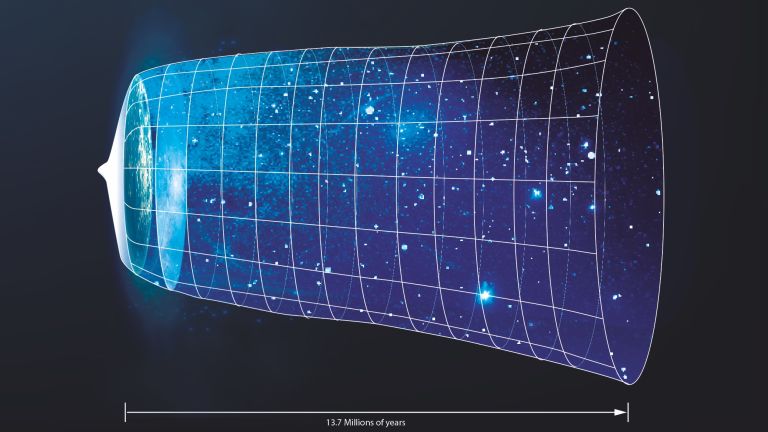
Without an internal clock, it would be impossible to adapt our lives to daily and seasonal fluctuations. Less obvious, but just as important for survival, is another type of time measurement that the nervous system handles: It recognizes which events occur more or less simultaneously. It constructs a “before” and an “after” from the signals of the sensory organs and distinguishes random occurrences from those linked as cause and effect.
Behind this lies one of evolution’s most ingenious “inventions”: the mechanism of synaptic integration. Put very simply, this helps us recognize what belongs together. Most neurons in the nervous system receive thousands of signals via their synapses and integrate them in such a way that a single output signal is produced: the Action potential Every second, the brain performs billions of such calculations. Signals sent by a single Neuron within a short period of time can be summed up just as easily as those originating from different nerve cells and arriving (almost) at the same time ▸ Communication of Cells. However, these stimuli are only transmitted if a certain threshold is exceeded. For example, events registered almost simultaneously by different sensory organs – such as a flash of lightning and the subsequent thunder, or a fall on the knee and the ensuing pain – can be “linked together.”
If the stimuli occur at too great a distance from one another or are not strong enough, they are “filtered out”: they apparently have nothing to do with one another. They are as insignificant as our hairstyle during a thunderstorm or the fact that we fell on a Tuesday. The situation is quite different when one experiences the same combination of stimuli multiple times. The nervous system can “adapt” to this by storing the experience and adjusting its reactions to it – in short: by learning.
Scientists researching our internal clock have now uncovered much of the inner workings and begun to understand their complex interplay. In doing so, they have gained unexpected insights that point far beyond the mechanistic. Their answers now even touch on philosophical questions regarding the nature of time. And they can also answer the mundane question of practical value with a clear conscience. After all, it was their work that laid the foundation for a better understanding and for future therapies of many neurological-psychiatric as well as organic diseases in which the internal clock has gone out of sync (see box).
Action potential
In excitable cells (e.g., neurons or muscle cells), very rapid changes in electrical potential occur across the cell membrane. This event is the basis for signal conduction along the axon of the nerve cell. The action potential continues along the cell membrane and, according to the all-or-nothing principle, only occurs when the cell has been sufficiently excited.
Neuron
A neuron is a specialized cell in the nervous system that is responsible for processing and transmitting information. It receives signals via its dendrites and transmits them via its axon. Transmission occurs electrically within the neuron and, between neurons, usually chemically via synapses.
Further reading
- Wilhelm, Klaus. Chronobiology: internal clocks in synch . Max-Plack-Gesellschaft (online). URL: https://www.mpg.de/10785637/chronobiology [Stand 25.3.2024]