When the Brain gets clogged with Waste
The brain is a highly active organ. To remove waste products from metabolism, it has a network of channels known as the glymphatic system. However, this system functions less effectively in old age and in cases of neurodegenerative diseases.
Scientific support: Prof. Dr. Petra Wahle
Published: 06.11.2024
Difficulty: easy
- The brain has its own waste disposal system, the glymphatic system, which extends along the blood vessels.
- According to current data, the brain’s waste removal system operates primarily at night.
- Sleep deprivation leads to an accumulation of misfolded proteins in the brain.
- Various neurodegenerative diseases, ranging from Dementia to Parkinson’s disease, could be linked to a dysfunctional glymphatic system.
- Impaired waste removal in the brain could influence the effects of a stroke or traumatic brain injury.
- However, the waste removal system does not operate in isolation but in conjunction with the lymphatic system, which cleanses all other organs and tissues.
Dementia
Dementia
Dementia is an acquired deficit of cognitive, social, motor, and emotional abilities. The most well-known form is Alzheimer's disease. "De mentia" means "without mind" in English.
stroke
Cerebral apoplexy
In a stroke, the brain or parts of it are no longer supplied with sufficient blood, which impairs the supply of oxygen and glucose. The most common cause is a blockage in an artery (ischemic stroke), less commonly a hemorrhage (hemorrhagic stroke). Typical symptoms include sudden visual disturbances, dizziness, paralysis, speech or sensory disturbances. Long-term consequences can include various sensory, motor, and cognitive impairments.
Well-shielded from the rest of the body, the brain floats in Cerebrospinal fluid beneath the skull. The Blood-brain barrier prevents dangerous pathogens and toxins from penetrating this sensitive organ and attacking it. For decades, neurologists believed that the brain, self-sufficient as it appears, could handle its own metabolic waste. After all, it also has its own immune system in the form of glial cells.
Yet there are seven grams of protein waste that must be cleared from the brain every day. Extrapolated, that would amount to a hefty two and a half kilograms per year. But how do these substances make their way out of the brain? And what if they don’t?
The rest of the human body’s organs are traversed by a lymphatic system that transports fluid away from the tissues, thereby regulating fluid balance and preventing conditions such as edema. Two to five liters of clear, pale yellow to milky fluid flow through the lymphatic vessels, flushing out not only excess tissue fluid but also toxins and pathogens from the spaces between cells and transporting them to the lymph nodes and into the bloodstream. For example, anyone who gets a tattoo will have colored lymph nodes shortly afterward, as some of the substances injected under the skin are transported there. Unlike blood, lymph fluid does not circulate. Instead, it is excreted via the kidneys and liver.
Cerebrospinal fluid
liquor cerebrospinalis
A clear fluid that fills the ventricular system and bathes the brain and spinal cord in the subarachnoid space, protecting them from impact. Three to five times a day, 100 to 160 ml of fluid is renewed by the choroid plexus. Certain diseases are reflected in the composition of the cerebrospinal fluid.
Blood-brain barrier
A selectively permeable membrane formed by cells in the walls of the capillary blood vessels in the brain. It protects the brain from harmful substances in the blood, but allows nutrients and oxygen to pass from the blood into the brain.
A dedicated waste disposal system for the brain
But how does the brain efficiently get rid of its waste products from cellular metabolism without any lymph at all? Researchers have asked themselves this question time and again in the past – including Danish neurobiologist Maiken Nedergaard from the University of Rochester in New York. To get to the bottom of this mystery, she injected a fluorescent tracer into the Cerebrospinal fluid of living mice. Using a multiphoton microscope, she observed how the tracer moved through the cerebrospinal fluid and within the brain. She was astonished: The dye in the cerebrospinal fluid rose through the spinal canal toward the brainstem and entered the brain via the fluid-filled space between the skull and the brain, known as the subarachnoid space. Alongside the blood vessels, a branched, previously unknown system of channels extends through the brain, mostly running parallel to the adjacent blood vessels. Later, the fluorescent substance disappeared again through channels that ran along the veins toward the trunk.
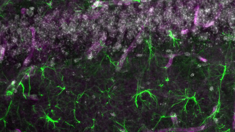
The water channels flank the blood vessels and are lined with astrocytes on the side facing the brain tissue. Astrocytes are star-shaped, branching nerve cells. Their extensions in the brain bear so-called end feet, and, as Nedergaard noted, these are dotted with numerous water-permeable pores. These consist of the protein aquaporin-4. Based on her findings, the neurobiologist concluded: The pores allow cerebrospinal fluid to branch off from the channel, which then flows into the spaces between the brain cells and continuously bathes them. In the process, the cerebrospinal fluid absorbs waste products.
Since the system resembles the lymphatic system, Nedergaard named it the “glymphatic system” in her 2012 publication. In doing so, she alluded to the well-known waste disposal system on the one hand and incorporated the term “glia” on the other – since astrocytes belong to this cell type.
Cerebrospinal fluid
liquor cerebrospinalis
A clear fluid that fills the ventricular system and bathes the brain and spinal cord in the subarachnoid space, protecting them from impact. Three to five times a day, 100 to 160 ml of fluid is renewed by the choroid plexus. Certain diseases are reflected in the composition of the cerebrospinal fluid.
Nighttime brain cleansing
Shortly thereafter, Nedergaard observed – again in mice – that the glymphatic system is primarily active at night. She watched as brain cells shrank during sleep, thereby increasing the water-flushed intercellular space – by as much as 60 percent. The neurobiologist then postulated that deeper brain regions are cleansed only at night. And when she added the protein Beta-amyloid – a component of the deposits in the brain associated with Alzheimer’s disease – to the mice’s cerebrospinal fluid, it reached the brain tissue more rapidly at night. In the journal Science in 2013, based on her experiments, she calculated that the inflow of Cerebrospinal fluid increased by 95 percent during sleep. Nedergaard concludes that “brain washing” operates at full speed at night.
In doing so, she puts forward a completely new possible explanation for neurodegenerative diseases such as Dementia and Parkinson’s disease: Whereas it was previously assumed that beta-amyloid and other misfolded and hyperphosphorylated proteins were broken down within the brain itself, Nedergaard now suggests that they are transported out via the glymphatic system. Could it be that the disposal of harmful proteins in neurodegenerative diseases no longer functions properly? Is the brain becoming cluttered with waste, causing nerve cells to die as a result?
There is now a wealth of evidence pointing in this direction. Many of the observations come from experiments on mice: for example, that harmful proteins such as beta-amyloids and tau in dementia, and the alpha-synuclein characteristic of Parkinson’s disease, accumulate in the brain when the glymphatic system is not functioning properly. This happens, for instance, when aquaporin-4 channels are blocked in mice due to a genetic mutation. According to Nedergaard’s research, the inflow of cerebrospinal fluid into the brain drops by 70 percent. In fact, in many neurological conditions – ranging from dementia to migraines and traumatic brain injuries – the glymphatic system, and specifically the aquaporin-4 channels, are altered.
Beta-amyloid
A peptide consisting of 36 to 42 amino acids that is considered the main component of senile plaques and is believed to be responsible for the development of Alzheimer's disease. The starting product is the amyloid precursor protein (APP). Certain enzymes in the cell membrane cut the precursor protein into peptides of various sizes. Amyloids consisting of 40 and 42 amino acids are found in senile plaques, with the 42-amino-acid product forming aggregates particularly quickly, at least in the Petri dish. The normal function of beta-amyloid has not yet been conclusively clarified.
Cerebrospinal fluid
liquor cerebrospinalis
A clear fluid that fills the ventricular system and bathes the brain and spinal cord in the subarachnoid space, protecting them from impact. Three to five times a day, 100 to 160 ml of fluid is renewed by the choroid plexus. Certain diseases are reflected in the composition of the cerebrospinal fluid.
Dementia
Dementia
Dementia is an acquired deficit of cognitive, social, motor, and emotional abilities. The most well-known form is Alzheimer's disease. "De mentia" means "without mind" in English.
Watching the brain’s waste disposal system at work
“The biggest limitation right now is that we can only observe the glymphatic system in humans indirectly,” laments Katerina Deike-Hoffmann, a neuroradiologist at the University Hospital of Bonn. “Our gold standard is a method so invasive that, to date, it has only been practiced by the research group led by radiologist Geir Ringstad and neurosurgeon Per Eide at the University Hospital in Oslo.” They inject the gadolinium-containing MRI contrast agent Gadubutrol into the Cerebrospinal fluid of volunteers at the level of the lumbar spine. This is a so-called off-label use: the drug is not approved for this application.
Therefore, both researchers initially limited their experiments to healthy individuals and investigated the effects of good sleep. After labeling their cerebrospinal fluid, the researchers kept seven participants awake overnight. Seventeen others went to bed at home as usual. The sensational findings earned both researchers a 2021 publication in the journal Brain: The contrast agent remained trapped in the brains of the sleep-deprived individuals, whereas it disappeared more quickly from the brains of the other participants. The varying degrees of waste product accumulation in the brain were evident in both groups, particularly in the cerebral Cortex – the region responsible for thinking, planning, and action – as well as in the emotional center, the limbic system, and the White matter (LINK). And although all participants subsequently had a night of normal sleep, the waste disposal system in the sleep-deprived group could no longer catch up. The experiments demonstrate for the first time in humans that the brain’s cleansing process occurs during sleep, Eide notes. And that lost sleep cannot be made up.
In line with this, the research group led by neurobiologist Nora Volkow of the National Institute of Health in Bethesda demonstrated as early as 2018 in 20 healthy subjects that a night without sleep increases the concentration of Beta-amyloid in certain regions of their brains. The areas affected were the right Hippocampus and the thalamus, which plays an important role in processing sensory input. The evidence of increased protein waste in the brain due to sleep deprivation aligns well with various longitudinal studies showing that people who sleep poorly have an increased risk of subsequently developing Dementia. Staying up all night is thus likely not only a consequence of the disease but also fuels it.
Cerebrospinal fluid
liquor cerebrospinalis
A clear fluid that fills the ventricular system and bathes the brain and spinal cord in the subarachnoid space, protecting them from impact. Three to five times a day, 100 to 160 ml of fluid is renewed by the choroid plexus. Certain diseases are reflected in the composition of the cerebrospinal fluid.
Cortex
cortex cerebri
Cortex refers to a collection of neurons, typically in the form of a thin surface. However, it usually refers to the cerebral cortex, the outermost layer of the cerebrum. It is 2.5 mm to 5 mm thick and rich in nerve cells. The cerebral cortex is heavily folded, comparable to a handkerchief in a cup. This creates numerous convolutions (gyri), fissures (fissurae), and sulci. Unfolded, the surface area of the cortex is approximately 1,800cm².
White matter
The white matter refers to the myelinated fibers of the nervous system that connect one neuron to another. The white color is caused by the myelin sheath surrounding the fibers.
Beta-amyloid
A peptide consisting of 36 to 42 amino acids that is considered the main component of senile plaques and is believed to be responsible for the development of Alzheimer's disease. The starting product is the amyloid precursor protein (APP). Certain enzymes in the cell membrane cut the precursor protein into peptides of various sizes. Amyloids consisting of 40 and 42 amino acids are found in senile plaques, with the 42-amino-acid product forming aggregates particularly quickly, at least in the Petri dish. The normal function of beta-amyloid has not yet been conclusively clarified.
Hippocampus
The hippocampus is the largest part of the archicortex and an area in the temporal lobe. It is also an important part of the limbic system. Functionally, it is involved in memory processes, but also in spatial orientation and learning. It comprises the subiculum, the dentate gyrus, and the Ammon's horn with its four fields CA1-CA4.
Changes in the structure of the hippocampus due to stress are associated with chronic pain. The hippocampus also plays an important role in the amplification of pain through anxiety.
Dementia
Dementia
Dementia is an acquired deficit of cognitive, social, motor, and emotional abilities. The most well-known form is Alzheimer's disease. "De mentia" means "without mind" in English.

Disrupted glymphatic system in various diseases
According to recent research, the glymphatic system also plays a role in many brain disorders and sometimes life-threatening brain injuries. In normal-pressure hydrocephalus, also known as age-related brain pressure, for example, the spaces containing Cerebrospinal fluid in the brain expand significantly. This causes fluid to accumulate in the head without a consistent increase in intracranial pressure. However, the brain tissue suffers, and the symptoms of the condition resemble early-stage Dementia. Biopsies of affected patients revealed a deficiency in aquaporin-4 channels.
This could explain why fluid accumulates in the head.
Other studies suggest that the extent to which the glymphatic system is affected could also influence the course of a traumatic brain injury and a stroke. Experiments in mice revealed that the inflow of cerebrospinal fluid plummeted following the occlusion of a cerebral artery – that is, a simulated stroke. When the artery spontaneously reopened, the flow of cerebrospinal fluid returned to normal.
Neuroscientists now also know that a significant portion of the long-term effects of a stroke is influenced by subsequent inflammation and the frequently occurring edema. According to Nedergaard, the edema originates directly from the glymphatic system. Shortly after the stroke, in her mouse experiments, the inflow of cerebrospinal fluid doubled and the space between brain cells expanded. A breakdown in the brain’s waste disposal system following a cerebral infarction could therefore lead to more severe physical and mental disabilities.
Cerebrospinal fluid
liquor cerebrospinalis
A clear fluid that fills the ventricular system and bathes the brain and spinal cord in the subarachnoid space, protecting them from impact. Three to five times a day, 100 to 160 ml of fluid is renewed by the choroid plexus. Certain diseases are reflected in the composition of the cerebrospinal fluid.
Dementia
Dementia
Dementia is an acquired deficit of cognitive, social, motor, and emotional abilities. The most well-known form is Alzheimer's disease. "De mentia" means "without mind" in English.
stroke
Cerebral apoplexy
In a stroke, the brain or parts of it are no longer supplied with sufficient blood, which impairs the supply of oxygen and glucose. The most common cause is a blockage in an artery (ischemic stroke), less commonly a hemorrhage (hemorrhagic stroke). Typical symptoms include sudden visual disturbances, dizziness, paralysis, speech or sensory disturbances. Long-term consequences can include various sensory, motor, and cognitive impairments.
The brain’s waste disposal system doesn’t work in isolation
With age, waste removal in the brain becomes increasingly less efficient. Pioneer Nedergaard pointed this out as early as 2014, when she noticed that mice up to 20 months old were less able to clear Beta-amyloid injected into the brain than their younger counterparts.
Recently, a Korean-American team succeeded in attributing the declining outflow of Cerebrospinal fluid in mice to the weakening lymphatic system in the nasopharynx. In doing so, they clearly demonstrate for the first time a connection between the well-known lymphatic system and the newly discovered glymphatic system.
This interaction is important: “The glymphatic system is intertwined with the well-known lymphatic system. It cannot be viewed in isolation,” emphasizes Swedish radiologist Geir Ringstad.
As evidence, he cites a study whose results were met with high expectations: In 2022, Ringstad’s research group was the first in the world to track how the brain’s cleansing process occurs in people with Dementia. He injected gadubutrol into the cerebrospinal fluid of 106 patients. At the same time, he monitored the levels of various disease-associated proteins in the patients’ blood. It could have been so simple: a better waste disposal system in the brain, less waste in the blood. But the picture that emerged for Ringstad is much more complicated: The levels of certain waste proteins, such as tau and another protein that indicates Neurodegeneration – the so-called light chain of the neurofilament protein – did indeed increase in blood plasma as brain cleansing decreased. But the performance of the glymphatic system was not the sole determining factor. Exchange between the cerebrospinal fluid and the blood was also necessary. And: Contrary to expectations, the beta-amyloid levels in the brains of those affected simply did not correlate with brain clearance capacity. The values showed no discernible connection. The authors agree: “Further research, primarily in humans, not in mice, is needed to understand how the glymphatic system truly functions and how it interacts with the lymphatic system.” It is hoped that, with further research, new therapeutic approaches for neurological diseases will emerge.
Beta-amyloid
A peptide consisting of 36 to 42 amino acids that is considered the main component of senile plaques and is believed to be responsible for the development of Alzheimer's disease. The starting product is the amyloid precursor protein (APP). Certain enzymes in the cell membrane cut the precursor protein into peptides of various sizes. Amyloids consisting of 40 and 42 amino acids are found in senile plaques, with the 42-amino-acid product forming aggregates particularly quickly, at least in the Petri dish. The normal function of beta-amyloid has not yet been conclusively clarified.
Cerebrospinal fluid
liquor cerebrospinalis
A clear fluid that fills the ventricular system and bathes the brain and spinal cord in the subarachnoid space, protecting them from impact. Three to five times a day, 100 to 160 ml of fluid is renewed by the choroid plexus. Certain diseases are reflected in the composition of the cerebrospinal fluid.
Dementia
Dementia
Dementia is an acquired deficit of cognitive, social, motor, and emotional abilities. The most well-known form is Alzheimer's disease. "De mentia" means "without mind" in English.
Neurodegeneration
Collective term for diseases in which nerve cells gradually lose their structure or function until they eventually die. In many cases, misfolded proteins are the trigger – such as certain forms of the proteins beta-amyloid and tau in the case of Alzheimer's disease. In other diseases, such as Parkinson's disease or Huntington's disease, proteins within the neurons are not broken down properly. As a result, toxic aggregates are deposited there, leading to the respective disease symptoms. While Huntington's disease is clearly genetic, in Parkinson's and Alzheimer's there appear to be certain gene variants that promote their development. None of these neurodegenerative diseases can be cured at present.
Further reading
- Nedergaard, M. et al: A Paravascular Pathway Facilitates CSF Flow Through the Brain Parenchyma and the Clearance of Interstitial Solutes, Including Amyloid β , Science Translational Medicine, 2012, 4, 147 ( zum Abstract )
- Nedergaard, M.: Garbage Truck of the Brain, Science, 2013, 340, 6140, 1529-1530 ( zum Volltext ).
- Nedergaard, M. et al: Sleep Drives Metabolite Clearance from the Adult Brain. Science, 2013, 342, 6156, 373-377 ( zum Volltext ).
- Eide, P. K. et al: Sleep deprivation impairs molecular clearance from the human brain, 2021, 144, 3, 863-874 ( zum Abstract )
- Volkow, N. et al: β -Amyloid accumulation in the human brain after one night of sleep deprivation, PNAS, 2018, 115, 17, 4483–4488 ( zum Volltext )
- Peng, S. et al: Aquaporin-4 in glymphatic system, and its implication for central nervous
- system disorders, Neurobiology of Disease, 2023, 179, 106035, ( zum Volltext )
- Nedergaard, M. et al: Impairment of paravascular clearance pathways in the aging brain, Annals of Neurology, 2014, 76, 6, 845-61 ( zum Abstract )
- Nedergaard, M. et al: Cerebrospinal fluid influx drives acute ischemic tissue swelling. Science, 2020, 13, 367, 6483, ( zum Volltext )
- Yoon, J.-H. et al: Nasopharyngeal lymphatic plexus is a hub for cerebrospinal fluid drainage, Nature, 2024, 625, 768–777 ( zum Volltext ).
- Eide, P. K. : Neurosurgery and the glymphatic system. Acta Neurologica, 2024, 166, 274, ( zum Volltext )
- Eide, P. K.: Plasma neurodegeneration biomarker concentrations associate with glymphatic and meningeal lymphatic measures in neurological disorders, Nature Communications, 14, 2084, ( zum Volltext )
