Hidden Channels
The path to discovering a waste disposal system in the brain was rocky and full of detours. We trace the journey and highlight the most important milestones.
Scientific support: Prof. Dr. Petra Wahle
Published: 06.11.2024
Difficulty: easy
- Paolo Mascagni discovered lymphatic vessels in the Dura mater as early as the 18th century, but his discovery was largely ignored and forgotten.
- Patricia Grady discovered in 1985 that Cerebrospinal fluid penetrates brain tissue, but her findings were not confirmed for a long time.
- Maiken Nedergaard described the glymphatic system in 2012 using two-photon microscopy; the system cleanses the brain during sleep.
- In 2023, Nedergaard described a fourth meninges that may separate clean cerebrospinal fluid from contaminated fluid and is important for the directed flow of cerebrospinal fluid.
Dura mater
The outermost of the three membranes covering the brain and spinal cord. Consists of connective tissue.
Cerebrospinal fluid
liquor cerebrospinalis
A clear fluid that fills the ventricular system and bathes the brain and spinal cord in the subarachnoid space, protecting them from impact. Three to five times a day, 100 to 160 ml of fluid is renewed by the choroid plexus. Certain diseases are reflected in the composition of the cerebrospinal fluid.
- The ventricles produce and distribute cerebrospinal fluid.
- The Cerebrospinal fluid is then transported along spaces that run parallel to the arteries in the brain.
- This transport is driven by the pulsating movements of the arteries.
- With the help of water channels located in the end feet of astrocytes, the cerebrospinal fluid is distributed into the brain tissue in the intercellular spaces.
- As the cerebrospinal fluid flows through the brain tissue, it absorbs metabolic byproducts and toxins.
- The fluid loaded with waste products is transported away through the spaces along the veins.
Cerebrospinal fluid
liquor cerebrospinalis
A clear fluid that fills the ventricular system and bathes the brain and spinal cord in the subarachnoid space, protecting them from impact. Three to five times a day, 100 to 160 ml of fluid is renewed by the choroid plexus. Certain diseases are reflected in the composition of the cerebrospinal fluid.
According to studies by Danish neuroscientist Maiken Nedergaard, waste removal in the brain is particularly effective during sleep. One possible reason for this is that neurons are thought to shrink during sleep, causing the intercellular spaces in the brain tissue to expand and thus facilitating the removal of waste. However, in 2024, researchers led by Nick Franks, Professor of Biophysics and Anesthesia at Imperial College London, reached the opposite conclusion: The clearance of substances in the brains of mice was not increased during sleep, but was actually significantly reduced. So do neurons actually shrink during sleep? And is waste removal truly more effective during slumber? That remains to be clarified. The differing results may be due to methodological differences: Maiken Nedergaard and her colleagues injected fluorescent markers into, among other places, the ventricles – cavities containing Cerebrospinal fluid The team led by Nick Franks, on the other hand, injected marker molecules directly into the brain.
Cerebrospinal fluid
liquor cerebrospinalis
A clear fluid that fills the ventricular system and bathes the brain and spinal cord in the subarachnoid space, protecting them from impact. Three to five times a day, 100 to 160 ml of fluid is renewed by the choroid plexus. Certain diseases are reflected in the composition of the cerebrospinal fluid.
In fact, the discovery should have found its way into anatomy textbooks on the brain long ago: The Italian anatomist Paolo Mascagni (1755–1815) had already identified lymphatic vessels in the Dura mater of the brain at the end of the 18th century. Due to technical difficulties, however, no one had been able to replicate Mascagni’s experiments and confirm the results. As a result, his discovery fell into oblivion again for more than two centuries. As recently as 2003, a renowned scientific journal remarked condescendingly: “Mascagni is so impressed by lymphatic vessels that he sees them everywhere, even where they do not exist, namely in the brain.”
In fact, until recently, researchers were convinced that the brain did not have its own lymphatic system. Outside the brain, the lymphatic system in the body ensures that waste is transported from the cells to the liver and kidneys, from where it finally leaves the body.
Researchers have now discovered lymphatic vessels in the meninges not only in animals but also in humans. In 2017, a team led by neurologist Daniel Reich from the U.S. National Institute of Neurological Disorders and stroke (NINDS) used Magnetic resonance imaging to observe how the human brain channels fluid through these vessels. The hypothesis is that this could serve as a waste disposal system.
We also know today that a structure exists in the brain that resembles the lymphatic system: the glymphatic system. The glymphatic system transports waste products within the brain. The path to its discovery was long and arduous. But after some detours, researchers finally found hidden channels in the brain.
An important milestone on the path to discovering the glymphatic system was an observation that, for a long time, could not be replicated. In the mid-1980s, the young neuroscientist Patricia Grady from the University of Maryland School of Medicine in Baltimore injected a tracer into the Cerebrospinal fluid of dogs or cats. Her surprising discovery: The cerebrospinal fluid penetrated the brain tissue along tiny channels that run parallel to the blood vessels.
This discovery was groundbreaking. It showed that cerebrospinal fluid enters the brain tissue itself, plays a role there in the transport and exchange of fluids, and may be involved in the removal of waste products. This transport along the blood vessels is a central component of the glymphatic system. But history repeated itself. As with the discovery of lymphatic vessels in the meninges in the 18th century, researchers were unable to reproduce Grady’s results. Consequently, it was mistakenly assumed that cerebrospinal fluid entered channels along the blood vessels only irregularly and in small amounts.
But admittedly, the glymphatic system doesn’t exactly make it easy for researchers to track it down. This is because the cleansing system is dynamic and heavily dependent on the flow of cerebrospinal fluid. It can therefore only be observed in the living brain.
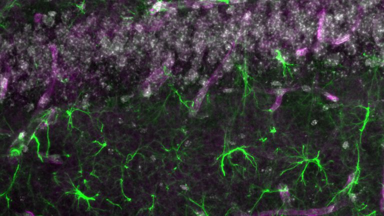
At least this time, it didn’t take over 200 years to finally confirm Patricia Grady’s findings. In 2012, Danish neuroscientist Maiken Nedergaard of the University of Rochester Medical Center used two-photon microscopy and fluorescent markers to track the movement of cerebrospinal fluid within the brain tissue of living mice. This revealed a network of channels running parallel to the arteries, along which cerebrospinal fluid penetrates the interstitial spaces of the brain tissue. The transport is driven by the pulse of the arteries. Studies in rodents also demonstrated that cerebrospinal fluid transport via the glymphatic system occurs rapidly – within minutes to hours – at least during sleep. Nedergaard and her colleagues named the system they discovered the glymphatic system.
Astrocytes, which surround the blood vessels and the interstitial space, are crucial for fluid exchange between the spaces along the blood vessels and the brain tissue. This is because the end feet of astrocytes contain water channel proteins (aquaporin 4) that channel cerebrospinal fluid into the brain tissue. In mice lacking the Gene for these water channel proteins, the inflow of cerebrospinal fluid into the brain is significantly reduced, and the removal of dissolved substances from the intercellular spaces in the brain tissue is reduced by 70 percent.
Dura mater
The outermost of the three membranes covering the brain and spinal cord. Consists of connective tissue.
stroke
Cerebral apoplexy
In a stroke, the brain or parts of it are no longer supplied with sufficient blood, which impairs the supply of oxygen and glucose. The most common cause is a blockage in an artery (ischemic stroke), less commonly a hemorrhage (hemorrhagic stroke). Typical symptoms include sudden visual disturbances, dizziness, paralysis, speech or sensory disturbances. Long-term consequences can include various sensory, motor, and cognitive impairments.
Magnetic resonance imaging
Magnetic resonance imaging scanner
A device used by medical professionals for magnetic resonance imaging (MRI). MRI is an imaging technique used to diagnose malformations in various tissues or organs of the body. This method is particularly effective for imaging parts of the body that contain a lot of water. Patients are placed in a tube (scanner) and exposed to a strong magnetic field. However, they are not exposed to X-rays or other forms of ionizing radiation.
Cerebrospinal fluid
liquor cerebrospinalis
A clear fluid that fills the ventricular system and bathes the brain and spinal cord in the subarachnoid space, protecting them from impact. Three to five times a day, 100 to 160 ml of fluid is renewed by the choroid plexus. Certain diseases are reflected in the composition of the cerebrospinal fluid.
Gene
Information unit on DNA. Specialized enzymes translate the core component of a gene into ribonucleic acid (RNA). While some ribonucleic acids perform important functions in the cell themselves, others specify the order in which the cell should assemble individual amino acids into a specific protein. The gene thus provides the code for this protein. In addition, a gene also includes regulatory elements on the DNA that ensure that the gene is read exactly when the cell or organism actually needs its product.
The fourth meninges
The glymphatic system, with its hidden channels, had thus been discovered. In 2023, another milestone followed that is likely to lead to a rewriting of the anatomical textbooks on the brain. Maiken Nedergaard discovered a fourth meningeal membrane in mice and humans, which today bears the complicated name “subarachnoid lymphatic membrane” (SLYM). Until this discovery, it was assumed that there were only three meninges protecting the brain: the dura mater, the outermost layer, followed by the arachnoid membrane, and the pia mater, the innermost layer.
The fourth meninges lies between the arachnoid and the pia mater and divides the so-called subarachnoid space. This space is filled with Cerebrospinal fluid As a fluid-filled buffer, it protects the brain from shocks and injuries and is also involved in the removal of waste products from the brain. The fourth meninges is as thin as tissue paper. Nevertheless, most proteins cannot pass through it, such as the protein beta-amyloid, whose deposits are associated with Alzheimer’s disease. In this respect, the fourth meninges may separate clean cerebrospinal fluid from contaminated fluid. In general, it appears to play an important role in the glymphatic system. A physically damaged fourth meninges impairs glymphatic flow.
Cerebrospinal fluid
liquor cerebrospinalis
A clear fluid that fills the ventricular system and bathes the brain and spinal cord in the subarachnoid space, protecting them from impact. Three to five times a day, 100 to 160 ml of fluid is renewed by the choroid plexus. Certain diseases are reflected in the composition of the cerebrospinal fluid.

Almost Escaped Discovery
The membrane had not been noticed before – partly because it dissolves when the brain is removed from the skull during an autopsy. Furthermore, it is too thin to show up in brain scans of living people. “In my view, there is strong evidence for a subdivision of the subarachnoid space, even if its exact organization is not yet entirely clear,” says neurosurgeon Per Kristian Eide of Oslo University Hospital.
The Nedergaard group’s characterization of the fourth meninges appears convincing. Reports on the membrane would certainly inspire other researchers and advance knowledge of the meninges. “The discovery of the fourth membrane is important because it reveals a subdivided subarachnoid space, which is significant for the directed flow of Cerebrospinal fluid within this space,” says Eide. This is important for the inflow to the glymphatic system.
Unlike Nedergaard’s study, much of the research on the glymphatic system has so far been conducted on animals, primarily mice. So how well has the waste disposal system been demonstrated in humans? “In my opinion, we have good evidence for the existence of a glymphatic system in humans,” says Per Kristian Eide. “However, it has not yet been possible to investigate all aspects of the glymphatic system in humans.” Studies by his own research group using human subjects and Magnetic resonance imaging provided evidence for various aspects: glymphatic inflow, the delayed clearance of glymphatic waste in certain diseases, and following sleep deprivation. “There is growing evidence for the existence of a human glymphatic system,” emphasizes Eide.
So it will be interesting to see when the discoveries surrounding the brain’s waste disposal system will finally find their way into neuroscience textbooks. Want to bet? It probably won’t take as long this time as it did for the discovery of lymphatic vessels in the meninges by Paolo Mascagni in the 18th century.
Cerebrospinal fluid
liquor cerebrospinalis
A clear fluid that fills the ventricular system and bathes the brain and spinal cord in the subarachnoid space, protecting them from impact. Three to five times a day, 100 to 160 ml of fluid is renewed by the choroid plexus. Certain diseases are reflected in the composition of the cerebrospinal fluid.
Magnetic resonance imaging
Magnetic resonance imaging scanner
A device used by medical professionals for magnetic resonance imaging (MRI). MRI is an imaging technique used to diagnose malformations in various tissues or organs of the body. This method is particularly effective for imaging parts of the body that contain a lot of water. Patients are placed in a tube (scanner) and exposed to a strong magnetic field. However, they are not exposed to X-rays or other forms of ionizing radiation.
Further reading
- Hablitz, L.M. et al.: Increased glymphatic influx is correlated with high EEG delta power and low heart rate in mice under anesthesia. In: Sci Adv 2019 Feb 27;5(2):eaav5447.
- Miao, A. et al.: Brain clearance is reduced during sleep and anesthesia. In: Nat Neurosci 2024 Jun;27(6):1046-1050.
EEG
An electroencephalogram, or EEG for short, is a recording of the brain's electrical activity (brain waves). Brain waves are measured on the surface of the head or using electrodes implanted in the brain itself. The time resolution is in the millisecond range, but the spatial resolution is very poor. The discoverer of electrical brain waves and EEG is the neurologist Hans Berger (1873−1941) from Jena.
