Balance: The vestibular Labyrinth
Published: 23.10.2025
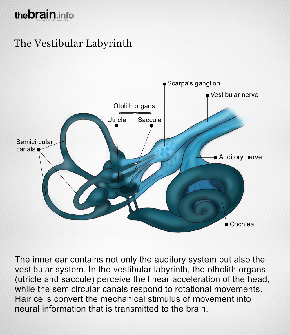
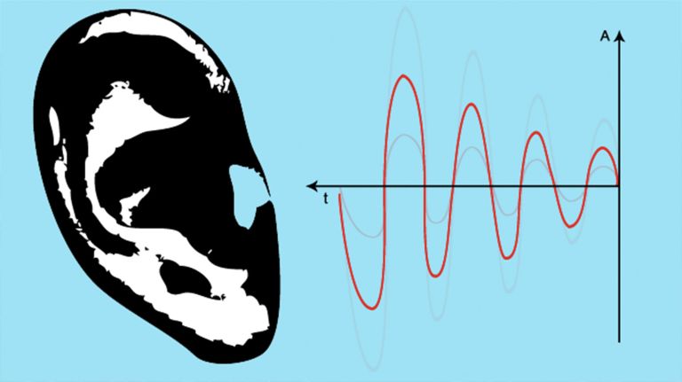
The Vestibular system ensures balance and stable vision. The Macula organs (utricle and saccule) register the linear acceleration of the head, while the Semicircular canals register its rotations.
Read more about this in our article ▸ A Labyrinth for Balance.
Vestibular system
Vestibular apparatus/Organon vestibulare/vestibular organ
The Vestibular system is part of the inner Ear. Its sensors are located in the Semicircular canals As part of the balance system, it detects circular movements (rotations), acceleration, and gravity.
macula lutea
The area of the Retina with the highest density of Photoreceptors. Due to this high "resolution," we see very sharply here. The diameter of the macula in humans is approximately 5 mm. The Fovea centralis is located in the center of the macula.
Semicircular canals
The three semicircular canals per ear are interconnected, fluid-filled tubes that are positioned almost at right angles to each other and belong to the balance organ in the inner ear (vestibular apparatus). They serve to register angular accelerations, i.e., rotational movements of the head.
Vestibular system
Vestibular apparatus/Organon vestibulare/vestibular organ
The vestibular system is part of the inner ear. Its sensors are located in the semicircular canals. As part of the balance system, it detects circular movements (rotations), acceleration, and gravity.
Ear
auris
The ear is not only the organ of hearing, but also of balance. A distinction is made between the outer ear with the auricle and external auditory canal, the middle ear with the eardrum and ossicles, and the actual hearing and balance organ, the inner ear with the cochlea and semicircular canals.
Semicircular canals
The three semicircular canals per ear are interconnected, fluid-filled tubes that are positioned almost at right angles to each other and belong to the balance organ in the inner ear (vestibular apparatus). They serve to register angular accelerations, i.e., rotational movements of the head.
Macula
macula lutea
The area of the retina with the highest density of photoreceptors. Due to this high "resolution," we see very sharply here. The diameter of the macula in humans is approximately 5 mm. The fovea centralis is located in the center of the macula.
Retina
The retina is the inner layer of the eye covered with pigment epithelium. The retina is characterized by an inverse (reversed) arrangement: light must first pass through several layers before it hits the photoreceptors (cones and rods). The signals from the photoreceptors are transmitted via the optic nerve to the processing areas of the brain. The reason for the inverse arrangement is the evolutionary development of the retina, which is a protrusion of the brain.
The retina is approximately 0.2 to 0.5 mm thick.
Photoreceptors
Photoreceptors are the light-sensitive cells of the retina; they convert light into electrical potentials. There are approximately 127 million photoreceptors in the retina, including seven million cones and 120 million rods.
Fovea centralis
The fovea centralis is located in the center of the macula and is the area of sharpest vision in birds and higher mammals. Its diameter in humans is approximately 1.5 mm. There are no rods in the fovea, only cones, which are interconnected to the ganglion cells in the central area of the fovea at a ratio of 1:1, resulting in very high "resolution."