Neuronal Burnout in the Hippocampus
Published: 10.05.2012
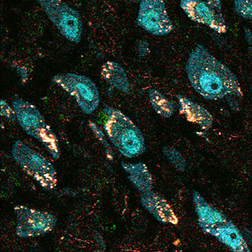
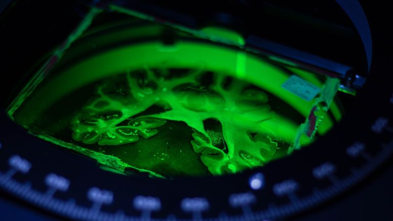
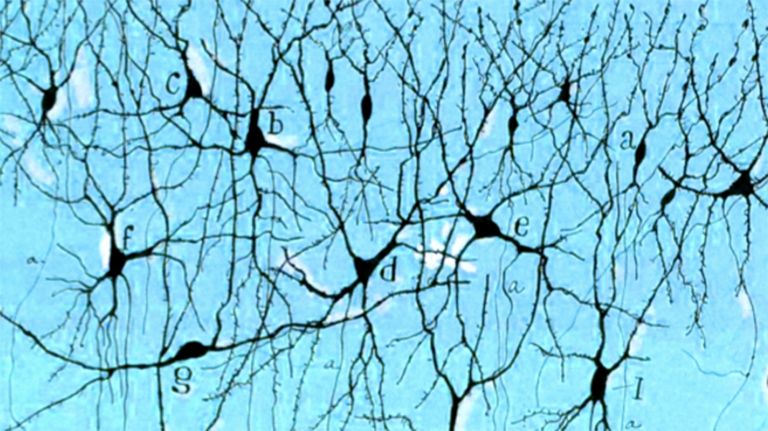
This is a cross-section of the hippocampus of an epilepsy patient for whom removal of the affected tissue was the only remaining treatment option. The neurons in this area produce proteins that have been altered due to cellular stress, which can pathologically alter communication between cells. In the image, the neuronal cell nuclei appear blue-green. The white dots surrounding them indicate the altered proteins.
For more information and exciting images on this topic, see our slideshow ▸ Communication under the microscope.
© Jochen Meier /MDC Berlin, 2010