
For an Enhanced Listening Experience – Optical Cochlear Implants generate more precise Nerve Signals than electrical Models
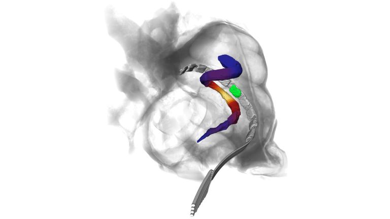
Researchers led by the University Medical Center Göttingen (UMG) have demonstrated in an animal model that optical cochlear implants generate nerve signals that are more easily distinguishable than those produced by conventional electrical implants. Nerve signals transmit auditory information – such as sound waves, pitch, and volume – to the brain, enabling a comprehensive hearing experience. In total, up to ten different light-emitting diodes (LEDs) were able to specifically trigger different nerve signals using very low levels of light energy. This underscores the potential of optical cochlear implants for improved hearing restoration. The results have been published in the journal “EMBO Molecular Medicine.”
Published: 05.11.2026
Today’s cochlear implants use electrical impulses to stimulate the Auditory nerve This enables many people who are deaf or have severe hearing loss to understand speech. The electrical current spreads through the conductive fluids of the cochlea, causing larger areas of the auditory nerve to be stimulated simultaneously and resulting in overlapping nerve signals. This limits the precision of the stimulation and makes it particularly difficult to understand speech in the presence of background noise. Modern cochlear implants have 12 to 24 “stimulation channels.” These channels allow the auditory nerve to be electrically stimulated, generating various nerve signals that are transmitted to the brain. In most cases, however, no more than ten channels can be distinguished. Results from previous animal studies have shown that the auditory nerve can be activated more precisely with light than with electrical impulses. This holds the promise of nerve signals that are more easily distinguishable from one another and, consequently, improved restoration of hearing.
A team led by Prof. Dr. Tobias Moser, Director of the Institute for Auditory Neuroscience at the University Medical Center Göttingen (UMG), genetically engineered the auditory nerve of Mongolian gerbils to express highly light-sensitive ion channels that can be selectively turned on and off like a “molecular light switch.” Even a small amount of light energy was then sufficient to stimulate the auditory nerve and thus trigger different nerve signals. These nerve signals are transmitted to the brain, where they are perceived as tones, sounds, or speech. The researchers were able to show that the nerve signals triggered by light could be distinguished from one another more clearly than with conventional stimulation using electrical impulses. “Our goal is to use optical cochlear implants to enable more distinguishable stimulation channels than with conventional electrical implants,” says Prof. Moser. “This would allow acoustic information to be transmitted to the auditory system in a more nuanced way. This could be of great benefit for restoring hearing in deaf and profoundly hard-of-hearing people, particularly for speech comprehension in the presence of background noise.”
The findings have been published in the journal “EMBO Molecular Medicine.”
Auditory nerve
nervus cochlearis
The hair cells of the organ of Corti stimulate neurons in the spiral ganglion, which is located in the cavity of the cochlea. Their axons form the auditory nerve, which transmits electrical impulses from the inner ear to the brain. Together with the vestibular nerve (nervus vestibularis), the auditory nerve forms the VIII cranial nerve.
How the hearing system reacts to light
The team investigated neural activity in a region of the Midbrain in Mongolian gerbils that is important for auditory processing. The researchers aimed to determine how information about different frequencies and intensities can be transmitted using light. To this end, they compared the neural signals triggered by light with those elicited by sounds or electrical cochlear implants.
The results showed that optical stimulation using LEDs could elicit neural signals that were roughly as strong as those triggered by electrical impulses or a sound with a sound pressure level of 51 decibels. “We were able to trigger distinct nerve signals using just a single LED,” says Niels Albrecht, a doctoral student at the Institute for Auditory Neuroscience at UMG and the study’s first author. “What is particularly interesting, however, is that the nerve signals triggered by different LEDs were easier to distinguish from one another than those produced by electrical stimulation – even when the LEDs were placed close together.”
Midbrain
mecencephalon
The midbrain is the uppermost section of the brain stem. Its regions are located around the aqueduct, a canal filled with cerebrospinal fluid. Prominent structures include the tectum, tegmentum, and substantia nigra.
Toward optogenetic hearing restoration
The study represents another important step toward the development of optical cochlear implants. Since optogenetic hearing restoration requires Gene therapy in addition to an implant, potential benefits and risks must be examined with particular care. The results from the animal model provide important insights in this regard and, at the same time, highlight which questions need to be addressed in future studies. These include, for example, whether stronger nerve signals can be achieved using more powerful light sources or even more light-sensitive ion channels. In addition, it must be investigated how more complex auditory information – such as speech – can be transmitted through the stimulation of multiple optical channels.
Gene
Information unit on DNA. Specialized enzymes translate the core component of a gene into ribonucleic acid (RNA). While some ribonucleic acids perform important functions in the cell themselves, others specify the order in which the cell should assemble individual amino acids into a specific protein. The gene thus provides the code for this protein. In addition, a gene also includes regulatory elements on the DNA that ensure that the gene is read exactly when the cell or organism actually needs its product.
Participating partners
In addition to UMG, researchers from the University of Göttingen, the German Primate Center – Leibniz Institute for Primate Research, the Max Planck Institute for Multidisciplinary Natural Sciences, and the University of Freiburg participated in the study. The work was supported, among others, by the Else Kröner Fresenius Center for Optogenetic Therapies (EKFZ OT), the Göttingen Cluster of Excellence “ “Multiscale Bioimaging: From Molecular Machines to Networks of Excitable Cells” (MBExC), and Collaborative Research Center 1690 “Disease Mechanisms and Functional Restoration of Sensory and Motor Systems.”
Original publication
Albrecht N, Koert E, Vavakou A et al. Multichannel optical cochlear implants enable spectrally distinct auditory activity. EMBO Molecular Medicine (2026). DOI: https://doi.org/10.1038/s44321-026-00541-2
