Types of Neurons
Published: 17.05.2012
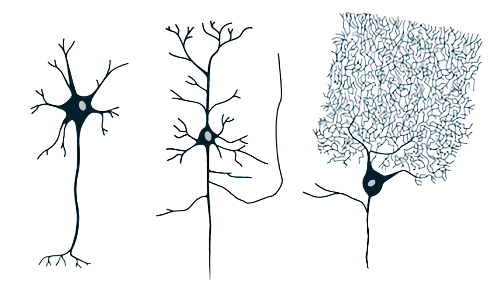
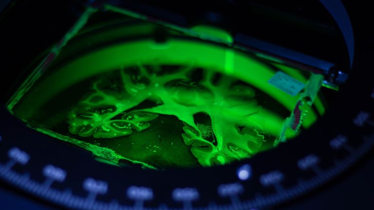
Multipolar neurons have different characteristics depending on their function. Here you can see a spinal Motor Neuron (left), a pyramidal cell in the Hippocampus (center), and a Purkinje cell in the Cerebellum (right). You can find more on this topic in our article by Leonie Seng. ▸ Cells: specialized Workers in the Brain
Motor neuron
Neurons that extend from the central nervous system to the muscles and control their activity.
The hippocampus is the largest part of the Archicortex and an area in the Temporal lobe It is also an important part of the Limbic system Functionally, it is involved in Memory processes, but also in spatial orientation and learning. It comprises the subiculum, the dentate gyrus, and the Ammon's horn with its four fields CA1-CA4.
Changes in the structure of the hippocampus due to stress are associated with chronic pain. The hippocampus also plays an important role in the amplification of pain through anxiety.
Purkinje cell
Purkinje cells are the main output cells of the Cerebellar cortex and central switching points of the Cerebellum. They have a dense, tree-like dendritic apparatus through which they receive information from thousands of parallel fibers and climbing fibers. Their axons are the only ones that extend out of the cerebellar Cortex and project onto the nuclei of the cerebellum, from where signals are transmitted to motor centers. Purkinje cells are among the largest cell types in the cerebellum.
Cerebellum
Cerebellum
The cerebellum is an important part of the brain, located at the back of the Brain stem and below the Occipital lobe It consists of two Cerebellar hemispheres covered by the cerebellar cortex and plays an important role in motor processes, among other things. It develops from the rhombencephalon.
Neuron
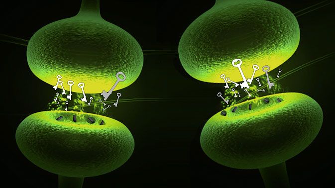
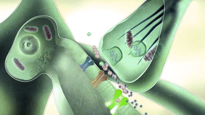
A neuron is a specialized cell in the nervous system that is responsible for processing and transmitting information. It receives signals via its dendrites and transmits them via its axon. Transmission occurs electrically within the neuron and, between neurons, usually chemically via synapses.
Hippocampus
The hippocampus is the largest part of the archicortex and an area in the temporal lobe. It is also an important part of the limbic system. Functionally, it is involved in memory processes, but also in spatial orientation and learning. It comprises the subiculum, the dentate gyrus, and the Ammon's horn with its four fields CA1-CA4.
Changes in the structure of the hippocampus due to stress are associated with chronic pain. The hippocampus also plays an important role in the amplification of pain through anxiety.
Archicortex
An ancient structure of the cerebrum in terms of evolutionary development, which, in contrast to the isocortex (also called the neocortex), has a three-layer structure. The archicortex mainly comprises the hippocampal structures.
Temporal lobe
Lobus temporalis
The temporal lobe is one of the four lobes of the cerebrum and is located laterally (on the side) at the bottom. It contains important areas such as the auditory cortex and parts of Wernicke's area, as well as areas for higher visual processing; deep within it lies the medial temporal lobe with structures such as the hippocampus.
Limbic system
The limbic system is a functional unit in the brain. It consists of interconnected structures, primarily in the cerebrum and diencephalon. The structures assigned to the system vary depending on the source, but the most important components are the hippocampus, amygdala, cingulate gyrus, septum, and mammillary bodies. The limbic system is involved in autonomic and visceral processes as well as in mechanisms of emotion, memory, and learning. Some authors mistakenly reduce the limbic system to the emotional world by referring to it as the "emotional brain."
Memory
Memory is a generic term for all types of information storage in the organism. In addition to pure retention, this also includes the absorption of information, its organization, and retrieval.
Ammon's horn
cornu ammonis
Part of the cerebrum, specifically the front end of the hippocampus. The Cornu Ammonis is divided into fields CA1 to CA4. It owes its name to its shape, which resembles the horn of an ammon sheep.
Cerebellar cortex
Cerebellar cortex
The cortex of the cerebellum, which, like that of the cerebrum, is composed of gray matter, or nerve cells. It consists of three layers and is highly folded, creating what are known as foliae, or leaves.
Cerebellum
Cerebellum
The cerebellum is an important part of the brain, located at the back of the brain stem and below the occipital lobe. It consists of two cerebellar hemispheres covered by the cerebellar cortex and plays an important role in motor processes, among other things. It develops from the rhombencephalon.
Cortex
cortex cerebri
Cortex refers to a collection of neurons, typically in the form of a thin surface. However, it usually refers to the cerebral cortex, the outermost layer of the cerebrum. It is 2.5 mm to 5 mm thick and rich in nerve cells. The cerebral cortex is heavily folded, comparable to a handkerchief in a cup. This creates numerous convolutions (gyri), fissures (fissurae), and sulci. Unfolded, the surface area of the cortex is approximately 1,800cm².
Brain stem
truncus cerebri
The "trunk" of the brain, to which all other brain structures are "attached," so to speak. From bottom to top, it comprises the medulla oblongata, the pons, and the mesencephalon. It transitions into the spinal cord below. It is a center for vital functions such as breathing and heartbeat and contains ascending and descending pathways between the cerebrum, cerebellum, and spinal cord.
Occipital lobe
lobus occipitalis
One of the four large lobes of the cerebral cortex. The occipital lobe lies above the cerebellum. It borders the parietal and temporal lobes at the front. The calcarine sulcus divides the occipital lobe into an upper and lower half, the cuneus and the lingual gyrus. Functionally, this area of the brain is responsible for the central processing of visual information – both the primary and secondary visual cortex are located in the occipital lobe.
Cerebellar hemispheres
Like the cerebrum, the cerebellum also has two hemispheres. The hemispheres are primarily responsible for finely tuned, purposeful movement control.