Somatotope Abbildung des Körpers
Published: 17.08.2011
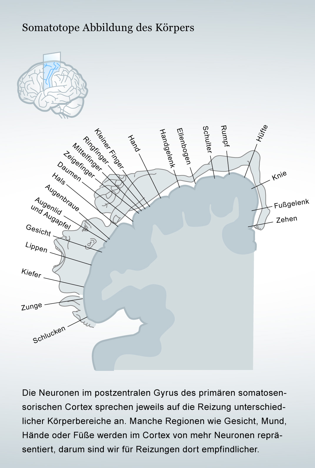
Zeichnet man die Körperbereiche ein, für die bestimmte Neuronengruppen im primären somatosensorischen Cortex zuständig sind, ergibt sich das Bild eines Homunculus, der ausgesteckt auf dem Cortex liegt. Mehr dazu wie bestimmte Gehirnareale unsere Körperteile kontrollieren, können Sie in dem Artikel von Kai Kupferschmiedt Das flexible Gehirn lernen.
Cortex
cortex cerebri
Cortex refers to a collection of neurons, typically in the form of a thin surface. However, it usually refers to the cerebral cortex, the outermost layer of the Cerebrum. It is 2.5 mm to 5 mm thick and rich in nerve cells. The cerebral cortex is heavily folded, comparable to a handkerchief in a cup. This creates numerous convolutions (gyri), fissures (fissurae), and sulci. Unfolded, the surface area of the cortex is approximately 1,800cm².
Cortex
cortex cerebri
Cortex refers to a collection of neurons, typically in the form of a thin surface. However, it usually refers to the cerebral cortex, the outermost layer of the cerebrum. It is 2.5 mm to 5 mm thick and rich in nerve cells. The cerebral cortex is heavily folded, comparable to a handkerchief in a cup. This creates numerous convolutions (gyri), fissures (fissurae), and sulci. Unfolded, the surface area of the cortex is approximately 1,800cm².
Cerebrum
telencephalon
The cerebrum comprises the cerebral cortex (gray matter), the nerve fibers (white matter), and the basal ganglia. It is the largest part of the brain. The cortex can be divided into four cortical areas: the temporal lobe, frontal lobe, occipital lobe, and parietal lobe.
Its functions include the coordination of perception, motivation, learning, and thinking.
