
Brain flexibly adapts to disruption
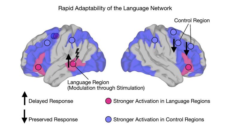
When an important area of the human brain involved in language processing is temporarily disrupted, other areas of the brain take over much of its work. Researchers at Leipzig University and the Max Planck Institute for Human Cognitive and Brain Sciences (MPI CBS) in Leipzig have shown how flexibly the brain can maintain performance even in the face of acute disruption, and how different brain networks work together in real time to achieve this. The researchers have just published their new study, which forms part of an ERC Consolidator Grant awarded to Professor Gesa Hartwigsen at Leipzig University and MPI CBS, in Nature Communications.
Published: 11.11.2026
“When semantic processing is disrupted, the brain flexibly recruits additional control regions that normally perform more general functions to support language processing. This allows it to maintain performance,” explains Hartwigsen. The study thus provides new insights into the brain’s ability to adapt rapidly and dynamically. Until now, research has primarily established which areas of the brain are involved in language processing and how the brain adapts over the longer term.What the researchers found particularly interesting was that they observed two opposing processes at the same time: while the language region directly affected by the disruption showed increased activity that was associated with poorer performance – reflected in slower response times – more distant control regions showed increased activity associated with better performance. This was associated with less impairment of language performance. “This suggests that disruption and compensation in the brain do not occur one after the other, but at the same time,” says Hartwigsen.In their study, the researchers combined two techniques that are typically used separately: transcranial magnetic stimulation (TMS), a method for stimulating the intact brain, and Functional magnetic resonance imaging (fMRI), a method for measuring brain activity. “Put simply, transcranial magnetic stimulation allowed us to temporarily and selectively interfere with processing in a small area of the brain. At the same time, functional Magnetic resonance imaging allowed us to observe what was happening across the entire brain,” explains Dr Anna-Lisa Schuler of MPI CBS, first author of the study.
These new findings could help scientists understand how we maintain cognitive performance, particularly in demanding everyday situations such as noisy environments. The study also provides initial indications of the processes that may be involved in the brain following sudden damage to regions involved in language – for example, after a stroke. In the long term, these findings could help inform new therapies aimed at restoring language and other cognitive functions following brain injury.The findings are based on studies of healthy young adults under laboratory conditions. Further research will be needed to determine the extent to which the findings apply to complex everyday situations, older people or people with neurological conditions.
Functional magnetic resonance imaging
Functional magnetic resonance imaging (fMRI) is a modification of MRI that allows brain activity to be measured indirectly via regional blood flow and oxygen consumption. BOLD (blood oxygen level dependent) contrast is often used, which exploits differences in the magnetic behavior of oxygen-rich and oxygen-poor blood. An increase in the BOLD signal indicates increased neural activity. fMRI provides good spatial resolution and allows detailed conclusions to be drawn about the activity of specific areas of the brain, while the temporal resolution is in the range of seconds.
Magnetic resonance imaging
Magnetic resonance imaging scanner
A device used by medical professionals for magnetic resonance imaging (MRI). MRI is an imaging technique used to diagnose malformations in various tissues or organs of the body. This method is particularly effective for imaging parts of the body that contain a lot of water. Patients are placed in a tube (scanner) and exposed to a strong magnetic field. However, they are not exposed to X-rays or other forms of ionizing radiation.
stroke
Cerebral apoplexy
In a stroke, the brain or parts of it are no longer supplied with sufficient blood, which impairs the supply of oxygen and glucose. The most common cause is a blockage in an artery (ischemic stroke), less commonly a hemorrhage (hemorrhagic stroke). Typical symptoms include sudden visual disturbances, dizziness, paralysis, speech or sensory disturbances. Long-term consequences can include various sensory, motor, and cognitive impairments.
Original publication
Anna-Lisa Schuler, Andrea Bruera & Gesa Hartwigsen; "Disrupting the Semantic Network Shifts Activity to Domain-General Brain Regions"; Nature Communcations
Source
