Search
-
- Glossary
Lateral geniculate body
The lateral geniculate nucleus is the section of the thalamus (the largest part of the diencephalon) where around 90% of the optic nerve axons terminate. It has a characteristic stratification into six cell layers. The nerve cells of the lateral geniculate nucleus send their projections to the visual cortex. Together with the medial geniculate nucleus, it forms the metathalamus.
-
- Glossary
Medial geniculate body
The medial geniculate body (medial geniculate nucleus) is a nucleus of the thalamus (the largest part of the diencephalon). As the central switching point of the auditory pathway, it transmits impulses from the inferior colliculus to the auditory radiation. Together with the lateral geniculate body, it forms the metathalamus.
-
- Percieve
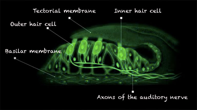
- Hearing
From Wiggling to the wonderful Variety of Sounds
It is a long way from purely mechanical vibrations to the world of sounds and tones.
17.10.2025
-
- Basics
- Anatomy
The Thalamus dorsalis
The structure is complex, the tasks are varied. It supports the senses, motor skills, and psyche.
28.11.2025
-
- News from the Institutes
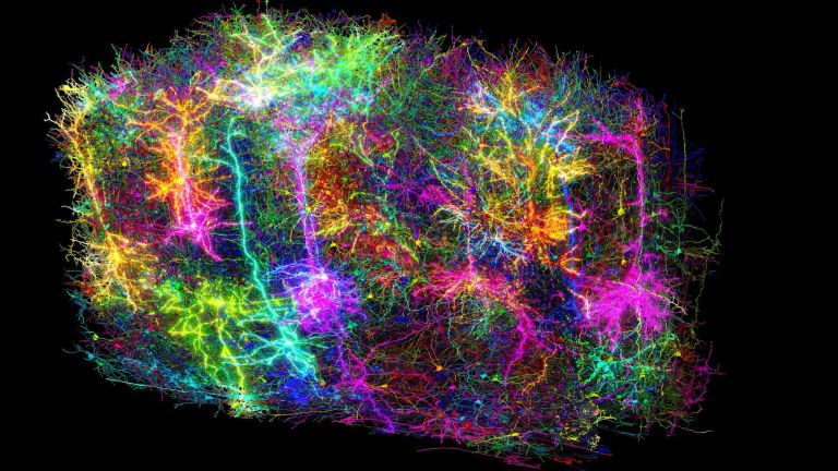
AI looks deeper into visual system
Artificial intelligence models provide insights to understand the processing of visual stimuli in the brain
10.04.2025
-
- Page
Glossary
15.03.2017