3D-Räume auf der Retina
Published: 30.08.2011
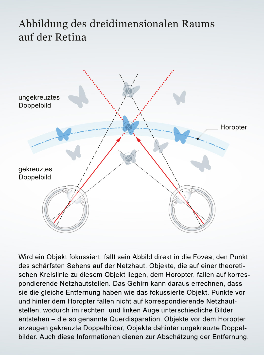
Dass wir den Raum dreidimensional erleben, verdanken wir komplexen Berechnungen des Gehirns – und dem Aufbau der Retina. Spannende Informationen zum Weiterlesen finden Sie in unserem Artikel Sehen in 3D von Hanna Drimalla.
The retina is the inner layer of the Eye covered with pigment epithelium. The retina is characterized by an inverse (reversed) arrangement: light must first pass through several layers before it hits the Photoreceptors (Cones and Rods). The signals from the photoreceptors are transmitted via the Optic nerve to the processing areas of the brain. The reason for the inverse arrangement is the evolutionary development of the retina, which is a protrusion of the brain.
The retina is approximately 0.2 to 0.5 mm thick.
Retina
The retina is the inner layer of the eye covered with pigment epithelium. The retina is characterized by an inverse (reversed) arrangement: light must first pass through several layers before it hits the photoreceptors (cones and rods). The signals from the photoreceptors are transmitted via the optic nerve to the processing areas of the brain. The reason for the inverse arrangement is the evolutionary development of the retina, which is a protrusion of the brain.
The retina is approximately 0.2 to 0.5 mm thick.
Eye
bulbus oculi
The eye is the sensory organ responsible for perceiving light stimuli – electromagnetic radiation within a specific frequency range. The light visible to humans lies in the range between 380 and 780 nanometers.
Photoreceptors
Photoreceptors are the light-sensitive cells of the retina; they convert light into electrical potentials. There are approximately 127 million photoreceptors in the retina, including seven million cones and 120 million rods.
Cones
The cones are a type of photoreceptor in the retina. The three different types of cones – S, M, and L – are each stimulated by short, medium, and long wavelengths of visible light, enabling color vision. They are highly concentrated in the fovea and enable sharp vision.
Rods
The rods are light-sensitive cells with high light sensitivity. They react even to weak light and are therefore responsible for scotopic vision, black-and-white vision, and vision at dusk. The rods are concentrated in the outer areas of the retina and therefore do not provide high visual acuity.
Optic nerve
nervus opticus
The axons (long fiber-like extensions) of the retinal ganglion cells form the optic nerve, which leaves the eye at the back of the optic disc. It comprises approximately one million axons and has a diameter of approximately seven millimeters.